Show PDB file:   
         Plain Text   HTML   (compressed file size)
QuickSearch:   
by PDB,NDB,UniProt,PROSITE Code or Search Term(s)  
(-)Asym.Unit - manually
(-)Asymmetric Unit
(-)Asym. Unit - sites
(-)Biological Unit 1
(-)Biol. Unit 1 - sites
(-)Biological Unit 2
(-)Biological Unit 3
(-)Biological Unit 4
(-)Biological Unit 5
(-)Biological Unit 6
collapse expand < >
Image Asym.Unit - manually
Asym.Unit - manually  (Jmol Viewer)
Image Asymmetric Unit
Asymmetric Unit  (Jmol Viewer)
Image Asym. Unit - sites
Asym. Unit - sites  (Jmol Viewer)
Image Biological Unit 1
Biological Unit 1  (Jmol Viewer)
Image Biol. Unit 1 - sites
Biol. Unit 1 - sites  (Jmol Viewer)
Image Biological Unit 2
Biological Unit 2  (Jmol Viewer)
Image Biological Unit 3
Biological Unit 3  (Jmol Viewer)
Image Biological Unit 4
Biological Unit 4  (Jmol Viewer)
Image Biological Unit 5
Biological Unit 5  (Jmol Viewer)
Image Biological Unit 6
Biological Unit 6  (Jmol Viewer)

(-) Description

Title :  CO-BOUND STRUCTURE OF BJFIXLH
 
Authors :  J. Key, K. Moffat
Date :  22 Sep 04  (Deposition) - 29 Mar 05  (Release) - 16 Nov 11  (Revision)
Method :  X-RAY DIFFRACTION
Resolution :  1.80
Chains :  Asym. Unit :  A,B
Biol. Unit 1:  A  (1x)
Biol. Unit 2:  B  (1x)
Biol. Unit 3:  A,B  (2x)
Biol. Unit 4:  A,B  (2x)
Biol. Unit 5:  A,B  (1x)
Biol. Unit 6:  A  (2x)
Keywords :  Pas Domain; Heme; Oxygen Sensor; Carbon Monoxide, Signaling Protein, Transferase (Keyword Search: [Gene Ontology, PubMed, Web (Google))
 
Reference :  J. Key, K. Moffat
Crystal Structures Of Deoxy And Co-Bound Bjfixlh Reveal Details Of Ligand Recognition And Signaling
Biochemistry V. 44 4627 2005
PubMed-ID: 15779889  |  Reference-DOI: 10.1021/BI047942R

(-) Compounds

Molecule 1 - SENSOR PROTEIN FIXL
    ChainsA, B
    EC Number2.7.3.-
    EngineeredYES
    Expression SystemESCHERICHIA COLI
    Expression System Taxid562
    Expression System Vector TypePLASMID
    FragmentHEME DOMAIN
    GeneFIXL
    Organism ScientificBRADYRHIZOBIUM JAPONICUM
    Organism Taxid375
    Other DetailsBOUND TO CARBON MONOXIDE
    SynonymBJFIXLH FIXL

 Structural Features

(-) Chains, Units

  12
Asymmetric Unit AB
Biological Unit 1 (1x)A 
Biological Unit 2 (1x) B
Biological Unit 3 (2x)AB
Biological Unit 4 (2x)AB
Biological Unit 5 (1x)AB
Biological Unit 6 (2x)A 

Summary Information (see also Sequences/Alignments below)

(-) Ligands, Modified Residues, Ions  (4, 7)

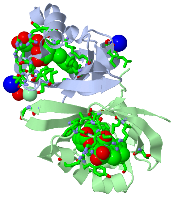
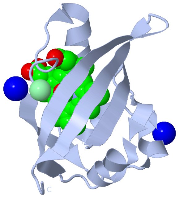
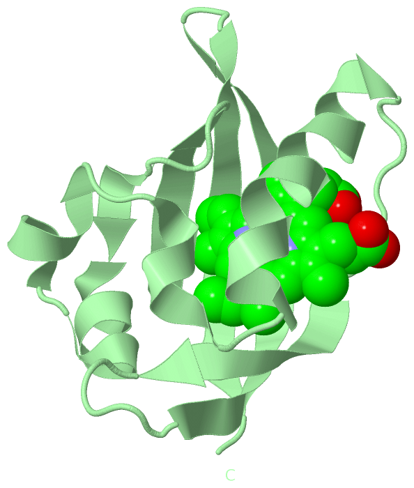
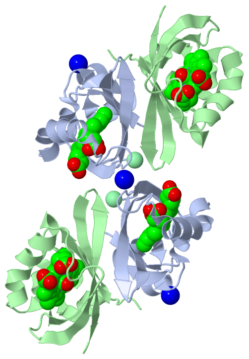
Asymmetric Unit (4, 7)
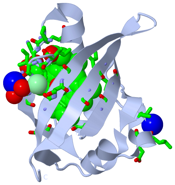
No.NameCountTypeFull Name
1CL1Ligand/IonCHLORIDE ION
2CMO2Ligand/IonCARBON MONOXIDE
3HEM2Ligand/IonPROTOPORPHYRIN IX CONTAINING FE
4NA2Ligand/IonSODIUM ION
Biological Unit 1 (2, 2)
No.NameCountTypeFull Name
1CL-1Ligand/IonCHLORIDE ION
2CMO1Ligand/IonCARBON MONOXIDE
3HEM1Ligand/IonPROTOPORPHYRIN IX CONTAINING FE
4NA-1Ligand/IonSODIUM ION
Biological Unit 2 (2, 2)
No.NameCountTypeFull Name
1CL-1Ligand/IonCHLORIDE ION
2CMO1Ligand/IonCARBON MONOXIDE
3HEM1Ligand/IonPROTOPORPHYRIN IX CONTAINING FE
4NA-1Ligand/IonSODIUM ION
Biological Unit 3 (2, 8)
No.NameCountTypeFull Name
1CL-1Ligand/IonCHLORIDE ION
2CMO4Ligand/IonCARBON MONOXIDE
3HEM4Ligand/IonPROTOPORPHYRIN IX CONTAINING FE
4NA-1Ligand/IonSODIUM ION
Biological Unit 4 (2, 8)
No.NameCountTypeFull Name
1CL-1Ligand/IonCHLORIDE ION
2CMO4Ligand/IonCARBON MONOXIDE
3HEM4Ligand/IonPROTOPORPHYRIN IX CONTAINING FE
4NA-1Ligand/IonSODIUM ION
Biological Unit 5 (2, 4)
No.NameCountTypeFull Name
1CL-1Ligand/IonCHLORIDE ION
2CMO2Ligand/IonCARBON MONOXIDE
3HEM2Ligand/IonPROTOPORPHYRIN IX CONTAINING FE
4NA-1Ligand/IonSODIUM ION
Biological Unit 6 (2, 4)
No.NameCountTypeFull Name
1CL-1Ligand/IonCHLORIDE ION
2CMO2Ligand/IonCARBON MONOXIDE
3HEM2Ligand/IonPROTOPORPHYRIN IX CONTAINING FE
4NA-1Ligand/IonSODIUM ION

(-) Sites  (7, 7)

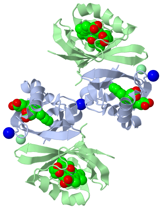
Asymmetric Unit (7, 7)
No.NameEvidenceResiduesDescription
1AC1SOFTWAREHOH A:82 , ILE A:218 , GLY A:219BINDING SITE FOR RESIDUE NA A 1001
2AC2SOFTWARELEU A:181 , GLU A:182 , ILE A:184BINDING SITE FOR RESIDUE NA A 1002
3AC3SOFTWAREHOH A:17 , GLY A:219 , SER A:237 , SER B:243 , GLY B:244BINDING SITE FOR RESIDUE CL A 1003
4AC4SOFTWAREHOH A:25 , ILE A:157 , ILE A:159 , LEU A:191 , MET A:192 , HIS A:200 , TYR A:203 , ARG A:206 , PRO A:213 , HIS A:214 , ILE A:215 , ILE A:216 , ARG A:220 , THR A:223 , LEU A:236 , PHE A:249 , GLY A:251 , CMO A:1720BINDING SITE FOR RESIDUE HEM A 719
5AC5SOFTWAREILE B:157 , ILE B:159 , LEU B:191 , MET B:192 , HIS B:200 , TYR B:203 , ARG B:206 , PRO B:213 , HIS B:214 , ILE B:215 , ILE B:216 , ARG B:220 , THR B:223 , LEU B:236 , PHE B:249 , GLY B:251 , CMO B:5720BINDING SITE FOR RESIDUE HEM B 719
6AC6SOFTWAREILE A:215 , LEU A:236 , ILE A:238 , HEM A:719BINDING SITE FOR RESIDUE CMO A 1720
7AC7SOFTWAREILE B:215 , LEU B:236 , ILE B:238 , HEM B:719BINDING SITE FOR RESIDUE CMO B 5720

(-) SS Bonds  (0, 0)

(no "SS Bond" information available for 1XJ4)

(-) Cis Peptide Bonds  (2, 2)
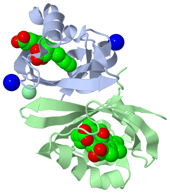
Asymmetric Unit
No.Residues
1Glu A:194 -Pro A:195
2Glu B:194 -Pro B:195

 Sequence-Structure Mapping

(-) SAPs(SNPs)/Variants  (0, 0)

(no "SAP(SNP)/Variant" information available for 1XJ4)

(-) PROSITE Motifs  (1, 2)

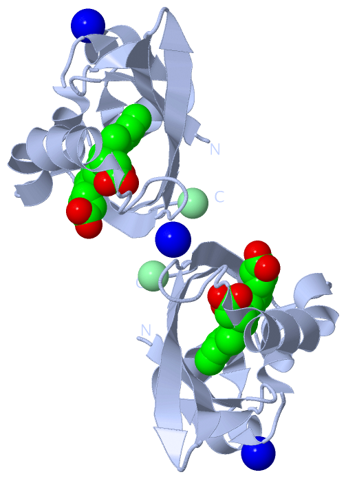
Asymmetric Unit (1, 2)
 PROSITEUniProtKBPDB
No.IDACDescriptionIDLocationCountLocation
1PACPS50113 PAC domain profile.FIXL_BRADU88-140
209-268
 
  2-
A:209-259
B:209-257
Biological Unit 1 (1, 1)
 PROSITEUniProtKBPDB
No.IDACDescriptionIDLocationCountLocation
1PACPS50113 PAC domain profile.FIXL_BRADU88-140
209-268
 
  1-
A:209-259
-
Biological Unit 2 (1, 1)
 PROSITEUniProtKBPDB
No.IDACDescriptionIDLocationCountLocation
1PACPS50113 PAC domain profile.FIXL_BRADU88-140
209-268
 
  1-
-
B:209-257
Biological Unit 3 (1, 4)
 PROSITEUniProtKBPDB
No.IDACDescriptionIDLocationCountLocation
1PACPS50113 PAC domain profile.FIXL_BRADU88-140
209-268
 
  4-
A:209-259
B:209-257
Biological Unit 4 (1, 4)
 PROSITEUniProtKBPDB
No.IDACDescriptionIDLocationCountLocation
1PACPS50113 PAC domain profile.FIXL_BRADU88-140
209-268
 
  4-
A:209-259
B:209-257
Biological Unit 5 (1, 2)
 PROSITEUniProtKBPDB
No.IDACDescriptionIDLocationCountLocation
1PACPS50113 PAC domain profile.FIXL_BRADU88-140
209-268
 
  2-
A:209-259
B:209-257
Biological Unit 6 (1, 2)
 PROSITEUniProtKBPDB
No.IDACDescriptionIDLocationCountLocation
1PACPS50113 PAC domain profile.FIXL_BRADU88-140
209-268
 
  2-
A:209-259
-

(-) Exons   (0, 0)

(no "Exon" information available for 1XJ4)

(-) Sequences/Alignments

Asymmetric Unit
   Reformat: Number of residues per line =  ('0' or empty: single-line sequence representation)
  Number of residues per labelling interval =   
  UniProt sequence: complete  aligned part    
   Show mapping: SCOP domains CATH domains Pfam domains Secondary structure (by author)
SAPs(SNPs) PROSITE motifs Exons
(details for a mapped element are shown in a popup box when the mouse pointer rests over it)
Chain A from PDB  Type:PROTEIN  Length:106
 aligned with FIXL_BRADU | P23222 from UniProtKB/Swiss-Prot  Length:505

    Alignment length:106
                                   163       173       183       193       203       213       223       233       243       253      
           FIXL_BRADU   154 DAMIVIDGHGIIQLFSTAAERLFGWSELEAIGQNVNILMPEPDRSRHDSYISRYRTTSDPHIIGIGRIVTGKRRDGTTFPMHLSIGEMQSGGEPYFTGFVRDLTEH 259
               SCOP domains d1xj4a_ A: Histidine kinase FixL heme domain                                                               SCOP domains
               CATH domains 1xj4A00 A:154-259  [code=3.30.450.20, no name defined]                                                     CATH domains
               Pfam domains ---------------------------------------------------------------------------------------------------------- Pfam domains
         Sec.struct. author .eeeeee....eeeehhhhhhhhh.hhhhhh..hhhhhh..hhhhhhhhhhhhhhhhh........eeeeee.....eeeeeeeeeeeee..eeeeeeeeee.... Sec.struct. author
                 SAPs(SNPs) ---------------------------------------------------------------------------------------------------------- SAPs(SNPs)
                    PROSITE -------------------------------------------------------PAC  PDB: A:209-259 UniProt: 209-268                PROSITE
                 Transcript ---------------------------------------------------------------------------------------------------------- Transcript
                 1xj4 A 154 DAMIVIDGHGIIQLFSTAAERLFGWSELEAIGQNVNILMPEPDRSRHDSYISRYRTTSDPHIIGIGRIVTGKRRDGTTFPMHLSIGEMQSGGEPYFTGFVRDLTEH 259
                                   163       173       183       193       203       213       223       233       243       253      

Chain B from PDB  Type:PROTEIN  Length:107
 aligned with FIXL_BRADU | P23222 from UniProtKB/Swiss-Prot  Length:505

    Alignment length:107
                                   160       170       180       190       200       210       220       230       240       250       
           FIXL_BRADU   151 TIPDAMIVIDGHGIIQLFSTAAERLFGWSELEAIGQNVNILMPEPDRSRHDSYISRYRTTSDPHIIGIGRIVTGKRRDGTTFPMHLSIGEMQSGGEPYFTGFVRDLT 257
               SCOP domains d1xj4b_ B: Histidine kinase FixL heme domain                                                                SCOP domains
               CATH domains 1xj4B00 B:151-257  [code=3.30.450.20, no name defined]                                                      CATH domains
               Pfam domains ----------------------------------------------------------------------------------------------------------- Pfam domains
         Sec.struct. author ....eeeeee....eeeehhhhhhhhh.hhhhhh..hhhhhh..hhhhhhhhhhhhhhh..........eeeeee.....eeeeeeeeeeeee..eeeeeeeeee.. Sec.struct. author
                 SAPs(SNPs) ----------------------------------------------------------------------------------------------------------- SAPs(SNPs)
                    PROSITE ----------------------------------------------------------PAC  PDB: B:209-257 UniProt: 209-268              PROSITE
                 Transcript ----------------------------------------------------------------------------------------------------------- Transcript
                 1xj4 B 151 TIPDAMIVIDGHGIIQLFSTAAERLFGWSELEAIGQNVNILMPEPDRSRHDSYISRYRTTSDPHIIGIGRIVTGKRRDGTTFPMHLSIGEMQSGGEPYFTGFVRDLT 257
                                   160       170       180       190       200       210       220       230       240       250       

   Legend:   → Mismatch (orange background)
  - → Gap (green background, '-', border residues have a numbering label)
    → Modified Residue (blue background, lower-case, 'x' indicates undefined single-letter code, labelled with number + name)
  x → Chemical Group (purple background, 'x', labelled with number + name, e.g. ACE or NH2)
  extra numbering lines below/above indicate numbering irregularities and modified residue names etc., number ends below/above '|'

 Classification and Annotation

(-) SCOP Domains  (1, 2)

Asymmetric Unit

(-) CATH Domains  (1, 2)

Asymmetric Unit
(-)
Class: Alpha Beta (26913)

(-) Pfam Domains  (0, 0)

(no "Pfam Domain" information available for 1XJ4)

(-) Gene Ontology  (16, 16)

Asymmetric Unit(hide GO term definitions)
Chain A,B   (FIXL_BRADU | P23222)
molecular function
    GO:0005524    ATP binding    Interacting selectively and non-covalently with ATP, adenosine 5'-triphosphate, a universally important coenzyme and enzyme regulator.
    GO:0016301    kinase activity    Catalysis of the transfer of a phosphate group, usually from ATP, to a substrate molecule.
    GO:0046872    metal ion binding    Interacting selectively and non-covalently with any metal ion.
    GO:0000166    nucleotide binding    Interacting selectively and non-covalently with a nucleotide, any compound consisting of a nucleoside that is esterified with (ortho)phosphate or an oligophosphate at any hydroxyl group on the ribose or deoxyribose.
    GO:0000155    phosphorelay sensor kinase activity    Catalysis of the phosphorylation of a histidine residue in response to detection of an extracellular signal such as a chemical ligand or change in environment, to initiate a change in cell state or activity. The two-component sensor is a histidine kinase that autophosphorylates a histidine residue in its active site. The phosphate is then transferred to an aspartate residue in a downstream response regulator, to trigger a response.
    GO:0004673    protein histidine kinase activity    Catalysis of the reaction: ATP + protein L-histidine = ADP + protein phospho-L-histidine.
    GO:0016740    transferase activity    Catalysis of the transfer of a group, e.g. a methyl group, glycosyl group, acyl group, phosphorus-containing, or other groups, from one compound (generally regarded as the donor) to another compound (generally regarded as the acceptor). Transferase is the systematic name for any enzyme of EC class 2.
    GO:0016772    transferase activity, transferring phosphorus-containing groups    Catalysis of the transfer of a phosphorus-containing group from one compound (donor) to another (acceptor).
biological process
    GO:0009399    nitrogen fixation    The process in which nitrogen is taken from its relatively inert molecular form (N2) in the atmosphere and converted into nitrogen compounds useful for other chemical processes, such as ammonia, nitrate and nitrogen dioxide.
    GO:0018106    peptidyl-histidine phosphorylation    The phosphorylation of peptidyl-histidine to form peptidyl-1'-phospho-L-histidine (otherwise known as tau-phosphohistidine, tele-phosphohistidine) or peptidyl-3'-phospho-L-histidine (otherwise known as pi-phosphohistidine, pros-phosphohistidine).
    GO:0000160    phosphorelay signal transduction system    A conserved series of molecular signals found in prokaryotes and eukaryotes; involves autophosphorylation of a histidine kinase and the transfer of the phosphate group to an aspartate that then acts as a phospho-donor to response regulator proteins.
    GO:0016310    phosphorylation    The process of introducing a phosphate group into a molecule, usually with the formation of a phosphoric ester, a phosphoric anhydride or a phosphoric amide.
    GO:0006355    regulation of transcription, DNA-templated    Any process that modulates the frequency, rate or extent of cellular DNA-templated transcription.
    GO:0007165    signal transduction    The cellular process in which a signal is conveyed to trigger a change in the activity or state of a cell. Signal transduction begins with reception of a signal (e.g. a ligand binding to a receptor or receptor activation by a stimulus such as light), or for signal transduction in the absence of ligand, signal-withdrawal or the activity of a constitutively active receptor. Signal transduction ends with regulation of a downstream cellular process, e.g. regulation of transcription or regulation of a metabolic process. Signal transduction covers signaling from receptors located on the surface of the cell and signaling via molecules located within the cell. For signaling between cells, signal transduction is restricted to events at and within the receiving cell.
    GO:0023014    signal transduction by protein phosphorylation    A process in which the transfer of one or more phosphate groups to a substrate transmits a signal to the phosphorylated substrate.
cellular component
    GO:0005622    intracellular    The living contents of a cell; the matter contained within (but not including) the plasma membrane, usually taken to exclude large vacuoles and masses of secretory or ingested material. In eukaryotes it includes the nucleus and cytoplasm.

 Visualization

(-) Interactive Views

Asymmetric Unit
  Complete Structure
    Jena3D(integrated viewing of ligand, site, SAP, PROSITE, SCOP information)
    WebMol | AstexViewer[tm]@PDBe
(Java Applets, require no local installation except for Java; loading may be slow)
    STRAP
(Java WebStart application, automatic local installation, requires Java; full application with system access!)
    RasMol
(require local installation)
    Molscript (VRML)
(requires installation of a VRML viewer; select preferred view via VRML and generate a mono or stereo PDF format file)
 
  Ligands, Modified Residues, Ions
    CL  [ RasMol | Jena3D ]  +environment [ RasMol | Jena3D ]
    CMO  [ RasMol | Jena3D ]  +environment [ RasMol | Jena3D ]
    HEM  [ RasMol | Jena3D ]  +environment [ RasMol | Jena3D ]
    NA  [ RasMol | Jena3D ]  +environment [ RasMol | Jena3D ]
 
  Sites
    AC1  [ RasMol ]  +environment [ RasMol ]
    AC2  [ RasMol ]  +environment [ RasMol ]
    AC3  [ RasMol ]  +environment [ RasMol ]
    AC4  [ RasMol ]  +environment [ RasMol ]
    AC5  [ RasMol ]  +environment [ RasMol ]
    AC6  [ RasMol ]  +environment [ RasMol ]
    AC7  [ RasMol ]  +environment [ RasMol ]
 
  Cis Peptide Bonds
    Glu A:194 - Pro A:195   [ RasMol ]  
    Glu B:194 - Pro B:195   [ RasMol ]  
 
Biological Units
  Complete Structure
    Biological Unit 1  [ Jena3D ]
    Biological Unit 2  [ Jena3D ]
    Biological Unit 3  [ Jena3D ]
    Biological Unit 4  [ Jena3D ]
    Biological Unit 5  [ Jena3D ]
    Biological Unit 6  [ Jena3D ]

(-) Still Images

Jmol
  protein: cartoon or spacefill or dots and stick; nucleic acid: cartoon and stick; ligands: spacefill; active site: stick
Molscript
  protein, nucleic acid: cartoon; ligands: spacefill; active site: ball and stick

 Databases and Analysis Tools

(-) Databases

Access by PDB/NDB ID
  1xj4
    Family and Domain InformationProDom | SYSTERS
    General Structural InformationGlycoscienceDB | MMDB | NDB | OCA | PDB | PDBe | PDBj | PDBsum | PDBWiki | PQS | PROTEOPEDIA
    Orientation in MembranesOPM
    Protein SurfaceSURFACE
    Secondary StructureDSSP (structure derived) | HSSP (homology derived)
    Structural GenomicsGeneCensus
    Structural NeighboursCE | VAST
    Structure ClassificationCATH | Dali | SCOP
    Validation and Original DataBMRB Data View | BMRB Restraints Grid | EDS | PROCHECK | RECOORD | WHAT_CHECK
 
Access by UniProt ID/Accession number
  FIXL_BRADU | P23222
    Comparative Protein Structure ModelsModBase
    Genomic InformationEnsembl
    Protein-protein InteractionDIP
    Sequence, Family and Domain InformationInterPro | Pfam | SMART | UniProtKB/SwissProt
 
Access by Enzyme Classificator   (EC Number)
  2.7.3.-
    General Enzyme InformationBRENDA | EC-PDB | Enzyme | IntEnz
    PathwayKEGG | MetaCyc
 
Access by Disease Identifier   (MIM ID)
  (no 'MIM ID' available)
    Disease InformationOMIM
 
Access by GenAge ID
  (no 'GenAge ID' available)
    Age Related InformationGenAge

(-) Analysis Tools

Access by PDB/NDB ID
    Domain InformationXDom
    Interatomic Contacts of Structural UnitsCSU
    Ligand-protein ContactsLPC
    Protein CavitiescastP
    Sequence and Secondary StructurePDBCartoon
    Structure AlignmentSTRAP(Java WebStart application, automatic local installation, requires Java; full application with system access!)
    Structure and Sequence BrowserSTING
 
Access by UniProt ID/Accession number
  FIXL_BRADU | P23222
    Protein Disorder PredictionDisEMBL | FoldIndex | GLOBPLOT (for more information see DisProt)

 Related Entries

(-) Entries Sharing at Least One Protein Chain (UniProt ID)

UniProtKB/Swiss-Prot
        FIXL_BRADU | P232221dp6 1dp8 1dp9 1drm 1lsv 1lsw 1lsx 1lt0 1xj2 1xj3 1xj6 1y28 2cmn 2owh 2owj 2vv6 2vv7 2vv8 4gcz

(-) Related Entries Specified in the PDB File

1xj2 1xj3 1xj6