Show PDB file:   
         Plain Text   HTML   (compressed file size)
QuickSearch:   
by PDB,NDB,UniProt,PROSITE Code or Search Term(s)  
(-)Asym./Biol. Unit - manually
(-)Asym./Biol. Unit
(-)Asym./Biol. Unit - sites
collapse expand < >
Image Asym./Biol. Unit - manually
Asym./Biol. Unit - manually  (Jmol Viewer)
Image Asym./Biol. Unit
Asym./Biol. Unit  (Jmol Viewer)
Image Asym./Biol. Unit - sites
Asym./Biol. Unit - sites  (Jmol Viewer)

(-) Description

Title :  STRUCTURE OF A PEPTIDE:N-GLYCANASE-RAD23 COMPLEX
 
Authors :  J. -H. Lee, J. M. Choi, C. Lee, K. J. Yi, Y. Cho
Date :  11 May 05  (Deposition) - 14 Jun 05  (Release) - 13 Jul 11  (Revision)
Method :  X-RAY DIFFRACTION
Resolution :  2.80
Chains :  Asym./Biol. Unit :  A,B,I
Keywords :  Hydrolase-Hydrolase Inhibitor Complex (Keyword Search: [Gene Ontology, PubMed, Web (Google))
 
Reference :  J. H. Lee, J. M. Choi, C. Lee, K. J. Yi, Y. Cho
Structure Of A Peptide:N-Glycanase-Rad23 Complex: Insight Into The Deglycosylation For Denatured Glycoproteins.
Proc. Natl. Acad. Sci. Usa V. 102 9144 2005
PubMed-ID: 15964983  |  Reference-DOI: 10.1073/PNAS.0502082102

(-) Compounds

Molecule 1
    ChainsA
    EC Number3.5.1.52
    EngineeredYES
    Expression SystemESCHERICHIA COLI BL21(DE3)
    Expression System StrainBL21(DE3)
    Expression System Taxid469008
    Expression System Vector TypePLASMID
    Organism CommonBAKER'S YEAST
    Organism ScientificSACCHAROMYCES CEREVISIAE
    Organism Taxid4932
 
Molecule 2 - UV EXCISION REPAIR PROTEIN RAD23
    ChainsB
    EngineeredYES
    Expression SystemESCHERICHIA COLI BL21(DE3)
    Expression System StrainBL21(DE3)
    Expression System Taxid469008
    Expression System Vector TypePLASMID
    FragmentXPC BINDING DOMAIN
    Organism CommonBAKER'S YEAST
    Organism ScientificSACCHAROMYCES CEREVISIAE
    Organism Taxid4932
 
Molecule 3 - PEPTIDE PHQ-VAL-ALA-ASP-CF0
    ChainsI
    EngineeredYES
    Other DetailsTHE PEPTIDE WAS CHEMICALLY SYNTHESIZED
    SyntheticYES

 Structural Features

(-) Chains, Units

  123
Asymmetric/Biological Unit ABI

Summary Information (see also Sequences/Alignments below)

(-) Ligands, Modified Residues, Ions  (3, 4)

Asymmetric/Biological Unit (3, 4)
No.NameCountTypeFull Name
1CF01Mod. Amino AcidFLUOROMETHANE
2SUC2Ligand/IonSUCROSE
3ZN1Ligand/IonZINC ION

(-) Sites  (4, 4)

Asymmetric Unit (4, 4)
No.NameEvidenceResiduesDescription
1AC1SOFTWARETRP A:123 , ASP A:217 , HIS A:218 , GLU A:238 , TRP A:251 , HOH A:1024 , ASP I:3BINDING SITE FOR RESIDUE SUC A1001
2AC2SOFTWARELYS A:117 , GLU A:238 , GLN A:239 , SER A:240 , GLN A:243 , ILE A:246BINDING SITE FOR RESIDUE SUC A1002
3AC3SOFTWARECYS A:129 , CYS A:132 , CYS A:165 , CYS A:168BINDING SITE FOR RESIDUE ZN A 999
4AC4SOFTWAREARG A:176 , ASN A:178 , ARG A:190 , CYS A:191 , GLY A:192 , GLU A:193 , ASP A:217 , SUC A:1001BINDING SITE FOR CHAIN I OF PEPTIDE PHQ-VAL-ALA- ASP-CF0

(-) SS Bonds  (0, 0)

(no "SS Bond" information available for 1X3Z)

(-) Cis Peptide Bonds  (0, 0)

(no "Cis Peptide Bond" information available for 1X3Z)

 Sequence-Structure Mapping

(-) SAPs(SNPs)/Variants  (0, 0)

(no "SAP(SNP)/Variant" information available for 1X3Z)

(-) PROSITE Motifs  (0, 0)

(no "PROSITE Motif" information available for 1X3Z)

(-) Exons   (2, 2)

Asymmetric/Biological Unit (2, 2)
 ENSEMBLUniProtKBPDB
No.Transcript IDExonExon IDGenome LocationLengthIDLocationLengthCountLocationLength
1.1YEL037C1YEL037C.1V:82603-814071197RAD23_YEAST1-3983981B:253-30957

2.1YPL096W1YPL096W.1XVI:366924-3680151092PNG1_YEAST1-3633631A:8-327320

(-) Sequences/Alignments

Asymmetric/Biological Unit
   Reformat: Number of residues per line =  ('0' or empty: single-line sequence representation)
  Number of residues per labelling interval =   
  UniProt sequence: complete  aligned part    
   Show mapping: SCOP domains CATH domains Pfam domains Secondary structure (by author)
SAPs(SNPs) PROSITE motifs Exons
(details for a mapped element are shown in a popup box when the mouse pointer rests over it)
Chain A from PDB  Type:PROTEIN  Length:320
 aligned with PNG1_YEAST | Q02890 from UniProtKB/Swiss-Prot  Length:363

    Alignment length:320
                                    17        27        37        47        57        67        77        87        97       107       117       127       137       147       157       167       177       187       197       207       217       227       237       247       257       267       277       287       297       307       317       327
           PNG1_YEAST     8 NNIDFDSIAKMLLIKYKDFILSKFKKAAPVENIRFQNLVHTNQFAQGVLGQSQHLCTVYDNPSWHSIVLETLDLDLIYKNVDKEFAKDGHAEGENIYTDYLVKELLRYFKQDFFKWCNKPDCNHCGQNTSENMTPLGSQGPNGEESKFNCGTVEIYKCNRCGNITRFPRYNDPIKLLETRKGRCGEWCNLFTLILKSFGLDVRYVWNREDHVWCEYFSNFLNRWVHVDSCEQSFDQPYIYSINWNKKMSYCIAFGKDGVVDVSKRYILQNELPRDQIKEEDLKFLCQFITKRLRYSLNDDEIYQLACRDEQEQIELIRGK 327
               SCOP domains d1x3za_ A: automated matches                                                                                                                                                                                                                                                                                                     SCOP domains
               CATH domains -------------------------------------------------------------------------------------------------------------------------------------------------------------------------------------------------------------------------------------------------------------------------------------------------------------------------------- CATH domains
               Pfam domains -------------------------------------------------------------------------------------------------------------------------------------------------------------------------------------------------------------Rad4-1x3zA01 A:213-308                                                                          ------------------- Pfam domains
         Sec.struct. author ....hhhhhhhhhhhhhhhhhhhh...hhhhhhhhhhhhhhhhhhhhhhhhhhhhhhhh.hhhhhhhhhhhhhhhhhhhhhhhhhhhh......hhhhhhhhhhhhhhhhhh..ee................eeeeeee...hhhhh..eeeeeeeee.....eeeeeee.hhhhhhhhheehhhhhhhhhhhhhhh....eeeeee...eeeeeeee....eeeeee....ee................eeeee..eeeehhhhhh..........hhhhhhhhhhhhhhhhhh..hhhhhhhhhhhhhhhhhhhhh.. Sec.struct. author
                 SAPs(SNPs) -------------------------------------------------------------------------------------------------------------------------------------------------------------------------------------------------------------------------------------------------------------------------------------------------------------------------------- SAPs(SNPs)
                    PROSITE -------------------------------------------------------------------------------------------------------------------------------------------------------------------------------------------------------------------------------------------------------------------------------------------------------------------------------- PROSITE
               Transcript 2 Exon 2.1  PDB: A:8-327 UniProt: 1-363 [INCOMPLETE]                                                                                                                                                                                                                                                                               Transcript 2
                 1x3z A   8 NNIDFDSIAKMLLIKYKDFILSKFKKAAPVENIRFQNLVHTNQFAQGVLGQSQHLCTVYDNPSWHSIVLETLDLDLIYKNVDKEFAKDGHAEGENIYTDYLVKELLRYFKQDFFKWCNKPDCNHCGQNTSENMTPLGSQGPNGEESKFNCGTVEIYKCNRCGNITRFPRYNDPIKLLETRKGRCGEWCNLFTLILKSFGLDVRYVWNREDHVWCEYFSNFLNRWVHVDSCEQSFDQPYIYSINWNKKMSYCIAFGKDGVVDVSKRYILQNELPRDQIKEEDLKFLCQFITKRLRYSLNDDEIYQLACRDEQEQIELIRGK 327
                                    17        27        37        47        57        67        77        87        97       107       117       127       137       147       157       167       177       187       197       207       217       227       237       247       257       267       277       287       297       307       317       327

Chain B from PDB  Type:PROTEIN  Length:57
 aligned with RAD23_YEAST | P32628 from UniProtKB/Swiss-Prot  Length:398

    Alignment length:57
                                   262       272       282       292       302       
          RAD23_YEAST   253 GSIGLTVEDLLSLRQVVSGNPEALAPLLENISARYPQLREHIMANPEVFVSMLLEAV 309
               SCOP domains d1x3zb_ B: automated matches                              SCOP domains
               CATH domains --------------------------------------------------------- CATH domains
               Pfam domains -----XPC-binding-1x3zB01 B:258-309                        Pfam domains
         Sec.struct. author .......hhhhhhhhhhhhh..hhhhhhhhhhh.hhhhhhhhhhhhhhhhhhhhhh. Sec.struct. author
                 SAPs(SNPs) --------------------------------------------------------- SAPs(SNPs)
                    PROSITE --------------------------------------------------------- PROSITE
               Transcript 1 Exon 1.1  PDB: B:253-309 UniProt: 1-398 [INCOMPLETE]      Transcript 1
                 1x3z B 253 GSIGLTVEDLLSLRQVVSGNPEALAPLLENISARYPQLREHIMANPEVFVSMLLEAV 309
                                   262       272       282       292       302       

Chain I from PDB  Type:PROTEIN  Length:4
                                    
               SCOP domains ---- SCOP domains
               CATH domains ---- CATH domains
               Pfam domains ---- Pfam domains
         Sec.struct. author .... Sec.struct. author
                 SAPs(SNPs) ---- SAPs(SNPs)
                    PROSITE ---- PROSITE
                 Transcript ---- Transcript
                 1x3z I   1 VADx   4
                               |
                               4-CF0

   Legend:   → Mismatch (orange background)
  - → Gap (green background, '-', border residues have a numbering label)
    → Modified Residue (blue background, lower-case, 'x' indicates undefined single-letter code, labelled with number + name)
  x → Chemical Group (purple background, 'x', labelled with number + name, e.g. ACE or NH2)
  extra numbering lines below/above indicate numbering irregularities and modified residue names etc., number ends below/above '|'

 Classification and Annotation

(-) SCOP Domains  (2, 2)

Asymmetric/Biological Unit

(-) CATH Domains  (0, 0)

(no "CATH Domain" information available for 1X3Z)

(-) Pfam Domains  (2, 2)

Asymmetric/Biological Unit

(-) Gene Ontology  (21, 25)

Asymmetric/Biological Unit(hide GO term definitions)
Chain A   (PNG1_YEAST | Q02890)
molecular function
    GO:0016787    hydrolase activity    Catalysis of the hydrolysis of various bonds, e.g. C-O, C-N, C-C, phosphoric anhydride bonds, etc. Hydrolase is the systematic name for any enzyme of EC class 3.
    GO:0046872    metal ion binding    Interacting selectively and non-covalently with any metal ion.
    GO:0000224    peptide-N4-(N-acetyl-beta-glucosaminyl)asparagine amidase activity    Catalysis of the reaction: 4-N-(N-acetyl-D-glucosaminyl)-protein + H2O = N-acetyl-beta-D-glucosaminylamine + peptide L-aspartate. This reaction is the hydrolysis of an N4-(acetyl-beta-D-glucosaminyl)asparagine residue in which the N-acetyl-D-glucosamine residue may be further glycosylated, to yield a (substituted) N-acetyl-beta-D-glucosaminylamine and the peptide containing an aspartic residue.
biological process
    GO:0006515    misfolded or incompletely synthesized protein catabolic process    The chemical reactions and pathways resulting in the breakdown of misfolded or attenuated proteins.
    GO:0006517    protein deglycosylation    The removal of sugar residues from a glycosylated protein.
cellular component
    GO:0005737    cytoplasm    All of the contents of a cell excluding the plasma membrane and nucleus, but including other subcellular structures.
    GO:0005829    cytosol    The part of the cytoplasm that does not contain organelles but which does contain other particulate matter, such as protein complexes.
    GO:0005634    nucleus    A membrane-bounded organelle of eukaryotic cells in which chromosomes are housed and replicated. In most cells, the nucleus contains all of the cell's chromosomes except the organellar chromosomes, and is the site of RNA synthesis and processing. In some species, or in specialized cell types, RNA metabolism or DNA replication may be absent.

Chain B   (RAD23_YEAST | P32628)
molecular function
    GO:0003684    damaged DNA binding    Interacting selectively and non-covalently with damaged DNA.
    GO:0000224    peptide-N4-(N-acetyl-beta-glucosaminyl)asparagine amidase activity    Catalysis of the reaction: 4-N-(N-acetyl-D-glucosaminyl)-protein + H2O = N-acetyl-beta-D-glucosaminylamine + peptide L-aspartate. This reaction is the hydrolysis of an N4-(acetyl-beta-D-glucosaminyl)asparagine residue in which the N-acetyl-D-glucosamine residue may be further glycosylated, to yield a (substituted) N-acetyl-beta-D-glucosaminylamine and the peptide containing an aspartic residue.
    GO:0070628    proteasome binding    Interacting selectively and non-covalently with a proteasome, a large multisubunit protein complex that catalyzes protein degradation.
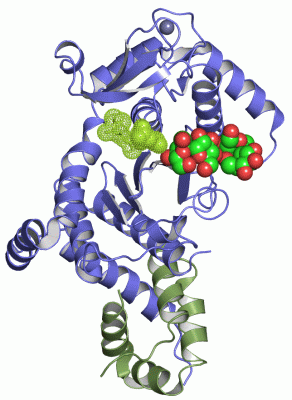
    GO:0005515    protein binding    Interacting selectively and non-covalently with any protein or protein complex (a complex of two or more proteins that may include other nonprotein molecules).
    GO:0030674    protein binding, bridging    The binding activity of a molecule that brings together two or more protein molecules, or a protein and another macromolecule or complex, through a selective, non-covalent, often stoichiometric interaction, permitting those molecules to function in a coordinated way.
    GO:0043130    ubiquitin binding    Interacting selectively and non-covalently with ubiquitin, a protein that when covalently bound to other cellular proteins marks them for proteolytic degradation.
biological process
    GO:0006281    DNA repair    The process of restoring DNA after damage. Genomes are subject to damage by chemical and physical agents in the environment (e.g. UV and ionizing radiations, chemical mutagens, fungal and bacterial toxins, etc.) and by free radicals or alkylating agents endogenously generated in metabolism. DNA is also damaged because of errors during its replication. A variety of different DNA repair pathways have been reported that include direct reversal, base excision repair, nucleotide excision repair, photoreactivation, bypass, double-strand break repair pathway, and mismatch repair pathway.
    GO:0006974    cellular response to DNA damage stimulus    Any process that results in a change in state or activity of a cell (in terms of movement, secretion, enzyme production, gene expression, etc.) as a result of a stimulus indicating damage to its DNA from environmental insults or errors during metabolism.
    GO:0000122    negative regulation of transcription from RNA polymerase II promoter    Any process that stops, prevents, or reduces the frequency, rate or extent of transcription from an RNA polymerase II promoter.
    GO:0006289    nucleotide-excision repair    A DNA repair process in which a small region of the strand surrounding the damage is removed from the DNA helix as an oligonucleotide. The small gap left in the DNA helix is filled in by the sequential action of DNA polymerase and DNA ligase. Nucleotide excision repair recognizes a wide range of substrates, including damage caused by UV irradiation (pyrimidine dimers and 6-4 photoproducts) and chemicals (intrastrand cross-links and bulky adducts).
    GO:0043161    proteasome-mediated ubiquitin-dependent protein catabolic process    The chemical reactions and pathways resulting in the breakdown of a protein or peptide by hydrolysis of its peptide bonds, initiated by the covalent attachment of ubiquitin, and mediated by the proteasome.
    GO:0006517    protein deglycosylation    The removal of sugar residues from a glycosylated protein.
    GO:0030433    ubiquitin-dependent ERAD pathway    The series of steps necessary to target endoplasmic reticulum (ER)-resident proteins for degradation by the cytoplasmic proteasome. Begins with recognition of the ER-resident protein, includes retrotranslocation (dislocation) of the protein from the ER to the cytosol, protein ubiquitination necessary for correct substrate transfer, transport of the protein to the proteasome, and ends with degradation of the protein by the cytoplasmic proteasome.
cellular component
    GO:0005737    cytoplasm    All of the contents of a cell excluding the plasma membrane and nucleus, but including other subcellular structures.
    GO:0000111    nucleotide-excision repair factor 2 complex    One of several protein complexes involved in nucleotide-excision repair; possesses damaged DNA binding activity. In S. cerevisiae, it is composed of Rad4p and Rad23p.
    GO:0005634    nucleus    A membrane-bounded organelle of eukaryotic cells in which chromosomes are housed and replicated. In most cells, the nucleus contains all of the cell's chromosomes except the organellar chromosomes, and is the site of RNA synthesis and processing. In some species, or in specialized cell types, RNA metabolism or DNA replication may be absent.
    GO:0000502    proteasome complex    A large multisubunit complex which catalyzes protein degradation, found in eukaryotes, archaea and some bacteria. In eukaryotes, this complex consists of the barrel shaped proteasome core complex and one or two associated proteins or complexes that act in regulating entry into or exit from the core.

 Visualization

(-) Interactive Views

Asymmetric/Biological Unit
  Complete Structure
    Jena3D(integrated viewing of ligand, site, SAP, PROSITE, SCOP information)
    WebMol | AstexViewer[tm]@PDBe
(Java Applets, require no local installation except for Java; loading may be slow)
    STRAP
(Java WebStart application, automatic local installation, requires Java; full application with system access!)
    RasMol
(require local installation)
    Molscript (VRML)
(requires installation of a VRML viewer; select preferred view via VRML and generate a mono or stereo PDF format file)
 
  Ligands, Modified Residues, Ions
    CF0  [ RasMol | Jena3D ]  +environment [ RasMol | Jena3D ]
    SUC  [ RasMol | Jena3D ]  +environment [ RasMol | Jena3D ]
    ZN  [ RasMol | Jena3D ]  +environment [ RasMol | Jena3D ]
 
  Sites
    AC1  [ RasMol ]  +environment [ RasMol ]
    AC2  [ RasMol ]  +environment [ RasMol ]
    AC3  [ RasMol ]  +environment [ RasMol ]
    AC4  [ RasMol ]  +environment [ RasMol ]
 
  Cis Peptide Bonds
(no "Cis Peptide Bonds" information available for 1x3z)
 

(-) Still Images

Jmol
  protein: cartoon or spacefill or dots and stick; nucleic acid: cartoon and stick; ligands: spacefill; active site: stick
Molscript
  protein, nucleic acid: cartoon; ligands: spacefill; active site: ball and stick

 Databases and Analysis Tools

(-) Databases

Access by PDB/NDB ID
  1x3z
    Family and Domain InformationProDom | SYSTERS
    General Structural InformationGlycoscienceDB | MMDB | NDB | OCA | PDB | PDBe | PDBj | PDBsum | PDBWiki | PQS | PROTEOPEDIA
    Orientation in MembranesOPM
    Protein SurfaceSURFACE
    Secondary StructureDSSP (structure derived) | HSSP (homology derived)
    Structural GenomicsGeneCensus
    Structural NeighboursCE | VAST
    Structure ClassificationCATH | Dali | SCOP
    Validation and Original DataBMRB Data View | BMRB Restraints Grid | EDS | PROCHECK | RECOORD | WHAT_CHECK
 
Access by UniProt ID/Accession number
  PNG1_YEAST | Q02890
    Comparative Protein Structure ModelsModBase
    Genomic InformationEnsembl
    Protein-protein InteractionDIP
    Sequence, Family and Domain InformationInterPro | Pfam | SMART | UniProtKB/SwissProt
  RAD23_YEAST | P32628
    Comparative Protein Structure ModelsModBase
    Genomic InformationEnsembl
    Protein-protein InteractionDIP
    Sequence, Family and Domain InformationInterPro | Pfam | SMART | UniProtKB/SwissProt
 
Access by Enzyme Classificator   (EC Number)
  3.5.1.52
    General Enzyme InformationBRENDA | EC-PDB | Enzyme | IntEnz
    PathwayKEGG | MetaCyc
 
Access by Disease Identifier   (MIM ID)
  (no 'MIM ID' available)
    Disease InformationOMIM
 
Access by GenAge ID
  (no 'GenAge ID' available)
    Age Related InformationGenAge

(-) Analysis Tools

Access by PDB/NDB ID
    Domain InformationXDom
    Interatomic Contacts of Structural UnitsCSU
    Ligand-protein ContactsLPC
    Protein CavitiescastP
    Sequence and Secondary StructurePDBCartoon
    Structure AlignmentSTRAP(Java WebStart application, automatic local installation, requires Java; full application with system access!)
    Structure and Sequence BrowserSTING
 
Access by UniProt ID/Accession number
  PNG1_YEAST | Q02890
    Protein Disorder PredictionDisEMBL | FoldIndex | GLOBPLOT (for more information see DisProt)
  RAD23_YEAST | P32628
    Protein Disorder PredictionDisEMBL | FoldIndex | GLOBPLOT (for more information see DisProt)

 Related Entries

(-) Entries Sharing at Least One Protein Chain (UniProt ID)

UniProtKB/Swiss-Prot
        PNG1_YEAST | Q028901x3w 3esw
        RAD23_YEAST | P326281x3w 2nbu 2nbw 2qsf 2qsg 2qsh 3esw 3m62 4yir

(-) Related Entries Specified in the PDB File

1x3w PEPTIDE:N-GLYCANASE-RAD23 COMPLEX