|
|
|
|
 Description
|
|
Compounds
|
||||||||||||||||||||||||||||||||||||||||
Chains, Units
Summary Information (see also Sequences/Alignments below) |
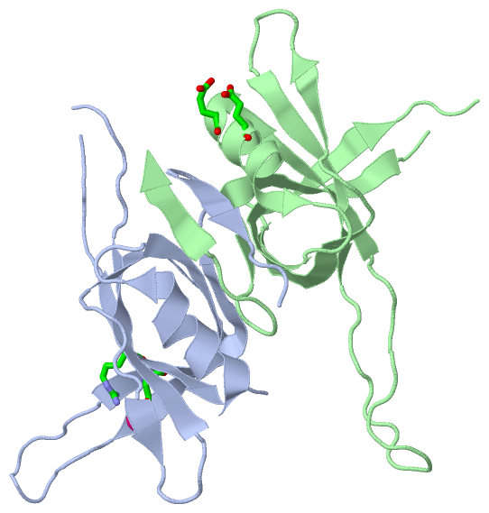
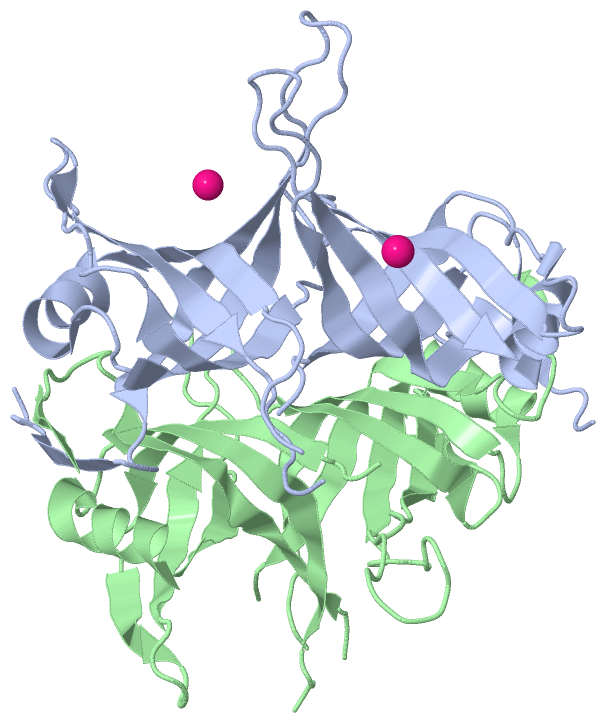
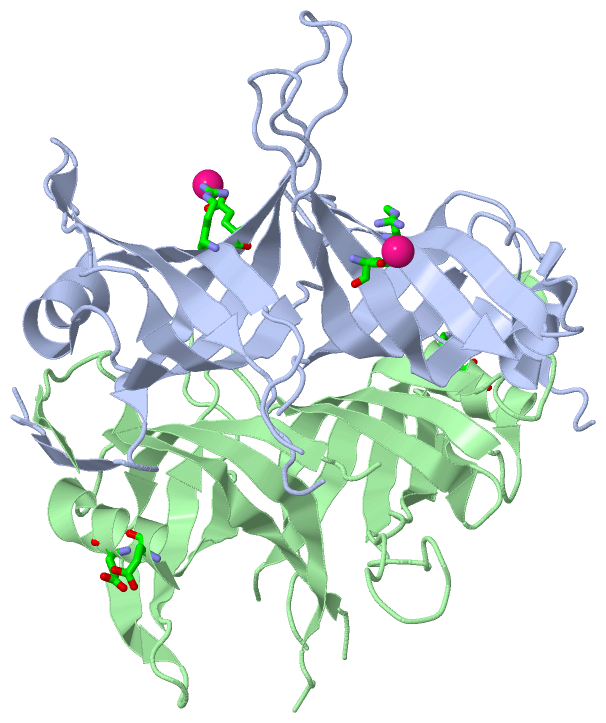
Ligands, Modified Residues, Ions (1, 1)
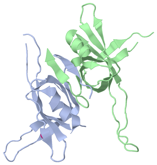
Asymmetric Unit (1, 1)
|
Sites (1, 1)
Asymmetric Unit (1, 1)
|
SS Bonds (0, 0)| (no "SS Bond" information available for 1X3G) |
Cis Peptide Bonds (0, 0)| (no "Cis Peptide Bond" information available for 1X3G) |
SAPs(SNPs)/Variants (0, 0)| (no "SAP(SNP)/Variant" information available for 1X3G) |
PROSITE Motifs (0, 0)| (no "PROSITE Motif" information available for 1X3G) |
Exons (0, 0)| (no "Exon" information available for 1X3G) |
Sequences/Alignments
Asymmetric UnitChain A from PDB Type:PROTEIN Length:114 aligned with SSB_MYCS2 | Q9AFI5 from UniProtKB/Swiss-Prot Length:165 Alignment length:118 12 22 32 42 52 62 72 82 92 102 112 SSB_MYCS2 3 GDTTITVVGNLTADPELRFTPSGAAVANFTVASTPRMFDRQSGEWKDGEALFLRCNIWREAAENVAESLTRGSRVIVTGRLKQRSFETREGEKRTVVEVEVDEIGPSLRYATAKVNKA 120 SCOP domains d1x3ga_ A: automated matches SCOP domains CATH domains 1x3gA00 A:3-120 Nucleic acid-binding pro teins CATH domains Pfam domains ---------------------------------------------------------------------------------------------------------------------- Pfam domains SAPs(SNPs) ---------------------------------------------------------------------------------------------------------------------- SAPs(SNPs) PROSITE ---------------------------------------------------------------------------------------------------------------------- PROSITE Transcript ---------------------------------------------------------------------------------------------------------------------- Transcript 1x3g A 3 GDTTITVVGNLTADPELRFTPSGAAVANFTVASTPRMFDR----WKDGEALFLRCNIWREAAENVAESLTRGSRVIVTGRLKQRSFETREGEKRTVVEVEVDEIGPSLRYATAKVNKA 120 12 22 32 42 | 52 62 72 82 92 102 112 42 47 Chain B from PDB Type:PROTEIN Length:115 aligned with SSB_MYCS2 | Q9AFI5 from UniProtKB/Swiss-Prot Length:165 Alignment length:118 12 22 32 42 52 62 72 82 92 102 112 SSB_MYCS2 3 GDTTITVVGNLTADPELRFTPSGAAVANFTVASTPRMFDRQSGEWKDGEALFLRCNIWREAAENVAESLTRGSRVIVTGRLKQRSFETREGEKRTVVEVEVDEIGPSLRYATAKVNKA 120 SCOP domains d1x3gb_ B: automated matches SCOP domains CATH domains 1x3gB00 B:3-120 Nucleic acid-binding proteins CATH domains Pfam domains (1) -SSB-1x3gB01 B:4-109 ----------- Pfam domains (1) Pfam domains (2) -SSB-1x3gB02 B:4-109 ----------- Pfam domains (2) SAPs(SNPs) ---------------------------------------------------------------------------------------------------------------------- SAPs(SNPs) PROSITE ---------------------------------------------------------------------------------------------------------------------- PROSITE Transcript ---------------------------------------------------------------------------------------------------------------------- Transcript 1x3g B 3 GDTTITVVGNLTADPELRFTPSGAAVANFTVASTPRMFDRQSGEWKDGEALFLRCNIWREAAENVAESLTRGSRVIVTGRLKQRSFETR---KRTVVEVEVDEIGPSLRYATAKVNKA 120 12 22 32 42 52 62 72 82 |- | 102 112 91 95
|
||||||||||||||||||||
SCOP Domains (1, 2)
Asymmetric Unit
 |
CATH Domains (1, 2)
Asymmetric Unit
|
Pfam Domains (1, 2)
Asymmetric Unit
|
Gene Ontology (3, 3)|
Asymmetric Unit(hide GO term definitions) Chain A,B (SSB_MYCS2 | Q9AFI5)
|
||||||||||||||||||||||||||||||
Interactive Views
|
||||||||||||||||||||||||||||||||||||||||||||||||||||||||||||||||||||||||||||||||||||||||||||||||||||||||||||||||||||||||||||||||||||||||
Still Images
|
||||||||||||||||
Databases
|
||||||||||||||||||||||||||||||||||||||||||||||||||||||||||||||||||||||||||||||||||||||||||||||||||||||||||||||||||||||||||||||||||||||||||||||||||||||||||||||||
Analysis Tools
|
|||||||||||||||||||||||||||||||||||||||||||||||||||||||||||||
Entries Sharing at Least One Protein Chain (UniProt ID)
Related Entries Specified in the PDB File
|
|