|
|
|
|
 Description
|
|
Compounds
|
||||||||||||||||||||||||||||||||||||||||||||
Chains, Units
Summary Information (see also Sequences/Alignments below) |
Ligands, Modified Residues, Ions (1, 1)
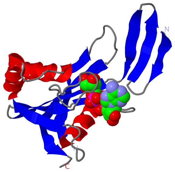
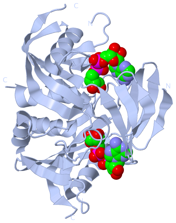
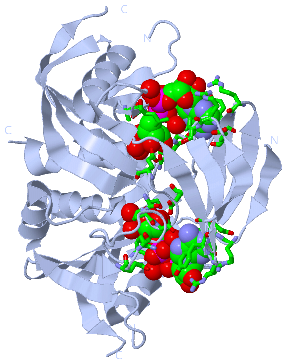
Asymmetric Unit (1, 1)
|
Sites (1, 1)
Asymmetric Unit (1, 1)
|
SS Bonds (0, 0)| (no "SS Bond" information available for 1V8L) |
Cis Peptide Bonds (0, 0)| (no "Cis Peptide Bond" information available for 1V8L) |
SAPs(SNPs)/Variants (0, 0)| (no "SAP(SNP)/Variant" information available for 1V8L) |
PROSITE Motifs (0, 0)| (no "PROSITE Motif" information available for 1V8L) |
Exons (0, 0)| (no "Exon" information available for 1V8L) |
Sequences/Alignments
Asymmetric UnitChain A from PDB Type:PROTEIN Length:157 aligned with Q84CU3_THETH | Q84CU3 from UniProtKB/TrEMBL Length:170 Alignment length:159 20 30 40 50 60 70 80 90 100 110 120 130 140 150 160 Q84CU3_THETH 11 RTYLYRGRILNLALEGRYEIVEHKPAVAVIALREGRMLFVRQMRPAVGLAPLEIPAGLIEPGEDPLEAARRELAEETGLSGDLTYLFSYFVSPGFTDEKTHVFLAENLKEVEAHPDEDEAIEVVWMRPEEALERHQRGEVEFSATGLVGVLYYHAFLRG 169 SCOP domains d1v8la_ A: ADP-ribose pyrophosphatase SCOP domains CATH domains 1v8lA00 A:11-169 Nucleoside Triphosphate Pyrophosphohydrolase CATH domains Pfam domains ----------------------NUDIX-1v8lA01 A:33-158 ----------- Pfam domains SAPs(SNPs) --------------------------------------------------------------------------------------------------------------------------------------------------------------- SAPs(SNPs) PROSITE --------------------------------------------------------------------------------------------------------------------------------------------------------------- PROSITE Transcript --------------------------------------------------------------------------------------------------------------------------------------------------------------- Transcript 1v8l A 11 RTYLYRGRILNLALEGRYEIVEHKPAVAVIALREGRMLFVRQMRPAVGLAPLEIPAGLIEPGEDPLEAARRELAEETGLSGDLTYLFSYFVSPGFTDEKTHVFLAENLKEVEA--DEDEAIEVVWMRPEEALERHQRGEVEFSATGLVGVLYYHAFLRG 169 20 30 40 50 60 70 80 90 100 110 120 | | 130 140 150 160 123 | 126
|
||||||||||||||||||||
SCOP Domains (1, 1)
Asymmetric Unit
 |
CATH Domains (1, 1)
Asymmetric Unit
|
Pfam Domains (1, 1)
Asymmetric Unit
|
Gene Ontology (2, 2)|
Asymmetric Unit(hide GO term definitions) Chain A (Q84CU3_THETH | Q84CU3)
|
||||||||||||||||||
Interactive Views
|
||||||||||||||||||||||||||||||||||||||||||||||||||||||||||||||||||||||||||||||||||||||||||||||||||||||||||||||||||||||||||||||||||||||||
Still Images
|
||||||||||||||||
Databases
|
||||||||||||||||||||||||||||||||||||||||||||||||||||||||||||||||||||||||||||||||||||||||||||||||||||||||||||||||||||||||||||||||||||||||||||||||||||||||||||||||
Analysis Tools
|
|||||||||||||||||||||||||||||||||||||||||||||||||||||||||||||
Entries Sharing at Least One Protein Chain (UniProt ID)
Related Entries Specified in the PDB File
|
|