Show PDB file:   
         Plain Text   HTML   (compressed file size)
QuickSearch:   
by PDB,NDB,UniProt,PROSITE Code or Search Term(s)  
(-)NMR Structure - model 1
(-)NMR Structure - all models
collapse expand < >
Image NMR Structure - model 1
NMR Structure - model 1  (Jmol Viewer)
Image NMR Structure - all models
NMR Structure - all models  (Jmol Viewer)

(-) Description

Title :  SOLUTION STRUCTURE OF PDZ DOMAIN OF MOUSE ALPHA-ACTININ-2 ASSOCIATED LIM PROTEIN
 
Authors :  N. Tochio, S. Koshiba, M. Inoue, T. Kigawa, S. Yokoyama, Riken Structural Genomics/Proteomics Initiative (Rsgi)
Date :  25 Nov 03  (Deposition) - 25 May 04  (Release) - 24 Feb 09  (Revision)
Method :  SOLUTION NMR
Resolution :  NOT APPLICABLE
Chains :  NMR Structure  :  A  (20x)
Keywords :  Pdz Domain, Cytoskeleton, Actin Binding, Structural Genomics, Riken Structural Genomics/Proteomics Initiative, Rsgi, Structural Protein (Keyword Search: [Gene Ontology, PubMed, Web (Google))
 
Reference :  N. Tochio, S. Koshiba, M. Inoue, T. Kigawa, S. Yokoyama
Solution Structure Of Pdz Domain Of Mouse Alpha-Actinin-2 Associated Lim Protein
To Be Published
PubMed: search
(for further references see the PDB file header)

(-) Compounds

Molecule 1 - PDZ AND LIM DOMAIN 3
    ChainsA
    EngineeredYES
    Expression System PlasmidP030506-95
    Expression System Vector TypePLASMID
    FragmentPDZ DOMAIN
    GeneRIKEN CDNA 6720456D19
    Organism CommonHOUSE MOUSE
    Organism ScientificMUS MUSCULUS
    Organism Taxid10090
    Other DetailsCELL-FREE PROTEIN SYNTHESIS

 Structural Features

(-) Chains, Units

  
NMR Structure (20x)

Summary Information (see also Sequences/Alignments below)

(-) Ligands, Modified Residues, Ions  (0, 0)

(no "Ligand,Modified Residues,Ions" information available for 1V5L)

(-) Sites  (0, 0)

(no "Site" information available for 1V5L)

(-) SS Bonds  (0, 0)

(no "SS Bond" information available for 1V5L)

(-) Cis Peptide Bonds  (2, 40)
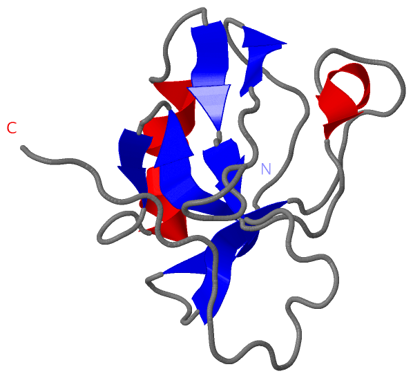
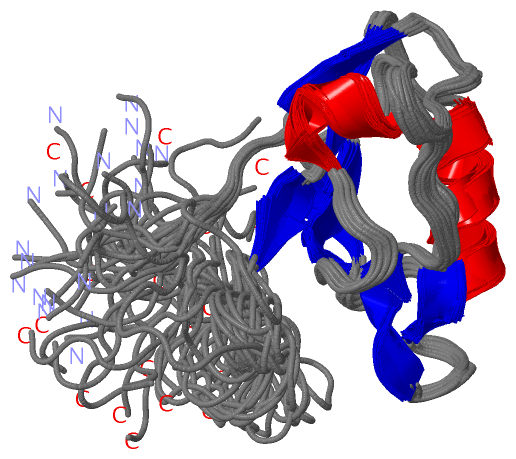
NMR Structure
No.ModelResidues
11, 2, 3, 4, 5, 6, 7, 8, 9, 10, 11, 12, 13, 14, 15, 16, 17, 18, 19, 20Gly A:13 -Pro A:14
21, 2, 3, 4, 5, 6, 7, 8, 9, 10, 11, 12, 13, 14, 15, 16, 17, 18, 19, 20Ala A:15 -Pro A:16

 Sequence-Structure Mapping

(-) SAPs(SNPs)/Variants  (0, 0)

(no "SAP(SNP)/Variant" information available for 1V5L)

(-) PROSITE Motifs  (1, 1)

NMR Structure (1, 1)
 PROSITEUniProtKBPDB
No.IDACDescriptionIDLocationCountLocation
1PDZPS50106 PDZ domain profile.PDLI3_MOUSE9-84  1A:13-88

(-) Exons   (0, 0)

(no "Exon" information available for 1V5L)

(-) Sequences/Alignments

NMR Structure
   Reformat: Number of residues per line =  ('0' or empty: single-line sequence representation)
  Number of residues per labelling interval =   
  UniProt sequence: complete  aligned part    
   Show mapping: SCOP domains CATH domains Pfam domains Secondary structure (by author)
SAPs(SNPs) PROSITE motifs Exons
(details for a mapped element are shown in a popup box when the mouse pointer rests over it)
Chain A from PDB  Type:PROTEIN  Length:103
 aligned with PDLI3_MOUSE | O70209 from UniProtKB/Swiss-Prot  Length:316

    Alignment length:124
                                1                                                                                                                       
                                |    6        16        26        36        46        56        66        76        86        96       106       116    
          PDLI3_MOUSE     - ----MPQNVVLPGPAPWGFRLSGGIDFNQPLVITRITPGSKAAAANLCPGDVILAIDGFGTESMTHADAQDRIKAASYQLCLKIDRAETRLWSPQVSEDGKAHPFKINLEAEPQEFKPIGTAHN 120
               SCOP domains d1v5la_ A: Alpha-actinin-2 associated LIM protein                                                                            SCOP domains
               CATH domains 1v5lA00 A:1-103  [code=2.30.42.10, no name defined]                                                                          CATH domains
               Pfam domains -------PDZ-1v5lA01 A:8-85                                                            --------------------------------------- Pfam domains
         Sec.struct. author .........eee.........eee....eeee.........hhhhh......eeee..ee....hhhhhhhhhh....eee.ee..............---------------------..... Sec.struct. author
                 SAPs(SNPs) ---------------------------------------------------------------------------------------------------------------------------- SAPs(SNPs)
                    PROSITE ------------PDZ  PDB: A:13-88 UniProt: 9-84                                             ------------------------------------ PROSITE
                 Transcript ---------------------------------------------------------------------------------------------------------------------------- Transcript
                 1v5l A   1 GSSGSSGNVVLPGPAPWGFRLSGGIDFNQPLVITRITPGSKAAAANLCPGDVILAIDGFGTESMTHADAQDRIKAASYQLCLKIDRAETRLWSPQVSS---------------------GPSSG 103
                                    10        20        30        40        50        60        70        80        90       | -         -        99    
                                                                                                                            98                    99    

   Legend:   → Mismatch (orange background)
  - → Gap (green background, '-', border residues have a numbering label)
    → Modified Residue (blue background, lower-case, 'x' indicates undefined single-letter code, labelled with number + name)
  x → Chemical Group (purple background, 'x', labelled with number + name, e.g. ACE or NH2)
  extra numbering lines below/above indicate numbering irregularities and modified residue names etc., number ends below/above '|'

 Classification and Annotation

(-) SCOP Domains  (1, 1)

NMR Structure

(-) CATH Domains  (1, 1)

NMR Structure
(-)
Class: Mainly Beta (13760)

(-) Pfam Domains  (1, 1)

NMR Structure
(-)
Clan: PDZ-like (184)

(-) Gene Ontology  (9, 9)

NMR Structure(hide GO term definitions)
Chain A   (PDLI3_MOUSE | O70209)
molecular function
    GO:0008092    cytoskeletal protein binding    Interacting selectively and non-covalently with any protein component of any cytoskeleton (actin, microtubule, or intermediate filament cytoskeleton).
    GO:0046872    metal ion binding    Interacting selectively and non-covalently with any metal ion.
    GO:0008307    structural constituent of muscle    The action of a molecule that contributes to the structural integrity of a muscle fiber.
    GO:0008270    zinc ion binding    Interacting selectively and non-covalently with zinc (Zn) ions.
biological process
    GO:0007015    actin filament organization    A process that is carried out at the cellular level which results in the assembly, arrangement of constituent parts, or disassembly of cytoskeletal structures comprising actin filaments. Includes processes that control the spatial distribution of actin filaments, such as organizing filaments into meshworks, bundles, or other structures, as by cross-linking.
    GO:0007507    heart development    The process whose specific outcome is the progression of the heart over time, from its formation to the mature structure. The heart is a hollow, muscular organ, which, by contracting rhythmically, keeps up the circulation of the blood.
cellular component
    GO:0030018    Z disc    Platelike region of a muscle sarcomere to which the plus ends of actin filaments are attached.
    GO:0015629    actin cytoskeleton    The part of the cytoskeleton (the internal framework of a cell) composed of actin and associated proteins. Includes actin cytoskeleton-associated complexes.
    GO:0005737    cytoplasm    All of the contents of a cell excluding the plasma membrane and nucleus, but including other subcellular structures.

 Visualization

(-) Interactive Views

NMR Structure
  Complete Structure
    Jena3D(integrated viewing of ligand, site, SAP, PROSITE, SCOP information)
    WebMol | AstexViewer[tm]@PDBe
(Java Applets, require no local installation except for Java; loading may be slow)
    STRAP
(Java WebStart application, automatic local installation, requires Java; full application with system access!)
    RasMol
(require local installation)
    Molscript (VRML)
(requires installation of a VRML viewer; select preferred view via VRML and generate a mono or stereo PDF format file)
 
  Ligands, Modified Residues, Ions
(no "Ligands, Modified Residues, Ions" information available for 1v5l)
 
  Sites
(no "Sites" information available for 1v5l)
 
  Cis Peptide Bonds
    Ala A:15 - Pro A:16   [ RasMol ]  
    Gly A:13 - Pro A:14   [ RasMol ]  
 

(-) Still Images

Jmol
  protein: cartoon or spacefill or dots and stick; nucleic acid: cartoon and stick; ligands: spacefill; active site: stick
Molscript
  protein, nucleic acid: cartoon; ligands: spacefill; active site: ball and stick

 Databases and Analysis Tools

(-) Databases

Access by PDB/NDB ID
  1v5l
    Family and Domain InformationProDom | SYSTERS
    General Structural InformationGlycoscienceDB | MMDB | NDB | OCA | PDB | PDBe | PDBj | PDBsum | PDBWiki | PQS | PROTEOPEDIA
    Orientation in MembranesOPM
    Protein SurfaceSURFACE
    Secondary StructureDSSP (structure derived) | HSSP (homology derived)
    Structural GenomicsGeneCensus
    Structural NeighboursCE | VAST
    Structure ClassificationCATH | Dali | SCOP
    Validation and Original DataBMRB Data View | BMRB Restraints Grid | EDS | PROCHECK | RECOORD | WHAT_CHECK
 
Access by UniProt ID/Accession number
  PDLI3_MOUSE | O70209
    Comparative Protein Structure ModelsModBase
    Genomic InformationEnsembl
    Protein-protein InteractionDIP
    Sequence, Family and Domain InformationInterPro | Pfam | SMART | UniProtKB/SwissProt
 
Access by Enzyme Classificator   (EC Number)
  (no 'Enzyme Classificator' available)
    General Enzyme InformationBRENDA | EC-PDB | Enzyme | IntEnz
    PathwayKEGG | MetaCyc
 
Access by Disease Identifier   (MIM ID)
  (no 'MIM ID' available)
    Disease InformationOMIM
 
Access by GenAge ID
  (no 'GenAge ID' available)
    Age Related InformationGenAge

(-) Analysis Tools

Access by PDB/NDB ID
    Domain InformationXDom
    Interatomic Contacts of Structural UnitsCSU
    Ligand-protein ContactsLPC
    Protein CavitiescastP
    Sequence and Secondary StructurePDBCartoon
    Structure AlignmentSTRAP(Java WebStart application, automatic local installation, requires Java; full application with system access!)
    Structure and Sequence BrowserSTING
 
Access by UniProt ID/Accession number
  PDLI3_MOUSE | O70209
    Protein Disorder PredictionDisEMBL | FoldIndex | GLOBPLOT (for more information see DisProt)

 Related Entries

(-) Entries Sharing at Least One Protein Chain (UniProt ID)

UniProtKB/Swiss-Prot
        PDLI3_MOUSE | O702091x64

(-) Related Entries Specified in the PDB File

(no "Related Entries Specified in the PDB File" available for 1V5L)