Show PDB file:   
         Plain Text   HTML   (compressed file size)
QuickSearch:   
by PDB,NDB,UniProt,PROSITE Code or Search Term(s)  
(-)Asym./Biol. Unit
(-)Asym./Biol. Unit - sites
collapse expand < >
Image Asym./Biol. Unit
Asym./Biol. Unit  (Jmol Viewer)
Image Asym./Biol. Unit - sites
Asym./Biol. Unit - sites  (Jmol Viewer)

(-) Description

Title :  STRUCTURE OF THE ENDOGLUCANASE CEL6 FROM MYCOBACTERIUM TUBERCULOSIS IN COMPLEX WITH CELLOBIOSE AT 1.75 ANGSTROM
 
Authors :  A. Varrot, S. Leydier, G. Pell, H. J. Gilbert, G. J. Davies
Date :  26 Sep 03  (Deposition) - 18 Nov 04  (Release) - 24 Feb 09  (Revision)
Method :  X-RAY DIFFRACTION
Resolution :  1.75
Chains :  Asym./Biol. Unit :  A
Keywords :  Hydrolase, Glycoside Hydrolase, Family 6 (Keyword Search: [Gene Ontology, PubMed, Web (Google))
 
Reference :  A. Varrot, S. Leydier, G. Pell, J. M. Macdonald, R. V. Stick, B. Henrissat, H. J. Gilbert, G. J. Davies
Mycobacterium Tuberculosis Strains Possess Functional Cellulases.
J. Biol. Chem. V. 280 20181 2005
PubMed-ID: 15824123  |  Reference-DOI: 10.1074/JBC.C500142200

(-) Compounds

Molecule 1 - PUTATIVE CELLULASE CEL6
    ChainsA
    EngineeredYES
    Expression SystemESCHERICHIA COLI
    Expression System PlasmidPET22B
    Expression System StrainORIGAMI
    Expression System Taxid562
    FragmentCATALYTIC DOMAIN, RESIDUES 88-380
    Organism ScientificMYCOBACTERIUM TUBERCULOSIS
    Organism Taxid83332
    Other DetailsSYNTHETIC GENE
    StrainH37RV
    SynonymENDOGLUCANASE, CELA

 Structural Features

(-) Chains, Units

  1
Asymmetric/Biological Unit A

Summary Information (see also Sequences/Alignments below)

(-) Ligands, Modified Residues, Ions  (5, 6)

Asymmetric/Biological Unit (5, 6)
No.NameCountTypeFull Name
11PG1Ligand/Ion2-(2-{2-[2-(2-METHOXY-ETHOXY)-ETHOXY]-ETHOXY}-ETHOXY)-ETHANOL
2ACT1Ligand/IonACETATE ION
3BGC2Ligand/IonBETA-D-GLUCOSE
4MHO1Mod. Amino AcidS-OXYMETHIONINE
5SO41Ligand/IonSULFATE ION

(-) Sites  (5, 5)

Asymmetric Unit (5, 5)
No.NameEvidenceResiduesDescription
1AC1SOFTWAREHIS A:247 , ASN A:277 , TRP A:319 , BGC A:1383 , HOH A:2212BINDING SITE FOR RESIDUE BGC A1382
2AC2SOFTWAREASP A:168 , ASP A:206 , MET A:210 , HIS A:247 , TRP A:250 , ASN A:277 , BGC A:1382 , HOH A:2108 , HOH A:2147 , HOH A:2207 , HOH A:2313 , HOH A:2314BINDING SITE FOR RESIDUE BGC A1383
3AC3SOFTWAREARG A:145 , TYR A:172 , THR A:355 , GLY A:357 , ARG A:358 , GLY A:359 , HOH A:2315BINDING SITE FOR RESIDUE SO4 A1384
4AC4SOFTWAREALA A:88 , ARG A:269 , HIS A:299 , HOH A:2316BINDING SITE FOR RESIDUE ACT A1385
5AC5SOFTWAREASN A:123 , THR A:124 , HOH A:2317BINDING SITE FOR RESIDUE 1PG A1386

(-) SS Bonds  (2, 2)

Asymmetric/Biological Unit
No.Residues
1A:169 -A:213
2A:320 -A:356

(-) Cis Peptide Bonds  (1, 1)

Asymmetric/Biological Unit
No.Residues
1Asn A:112 -Pro A:113

 Sequence-Structure Mapping

(-) SAPs(SNPs)/Variants  (0, 0)

(no "SAP(SNP)/Variant" information available for 1UP0)

(-) PROSITE Motifs  (0, 0)

(no "PROSITE Motif" information available for 1UP0)

(-) Exons   (0, 0)

(no "Exon" information available for 1UP0)

(-) Sequences/Alignments

Asymmetric/Biological Unit
   Reformat: Number of residues per line =  ('0' or empty: single-line sequence representation)
  Number of residues per labelling interval =   
  UniProt sequence: complete  aligned part    
   Show mapping: SCOP domains CATH domains Pfam domains Secondary structure (by author)
SAPs(SNPs) PROSITE motifs Exons
(details for a mapped element are shown in a popup box when the mouse pointer rests over it)
Chain A from PDB  Type:PROTEIN  Length:293
 aligned with Q79G13_MYCTU | Q79G13 from UniProtKB/TrEMBL  Length:380

    Alignment length:293
                                    97       107       117       127       137       147       157       167       177       187       197       207       217       227       237       247       257       267       277       287       297       307       317       327       337       347       357       367       377   
         Q79G13_MYCTU    88 ANPLAGKPFYVDPASAAMVAARNANPPNAELTSVANTPQSYWLDQAFPPATVGGTVARYTGAAQAAGAMPVLTLYGIPHRDCGSYASGGFATGTDYRGWIDAVASGLGSSPATIIVEPDALAMADCLSPDQRQERFDLVRYAVDTLTRDPAAAVYVDAGHSRWLSAEAMAARLNDVGVGRARGFSLNVSNFYTTDEEIGYGEAISGLTNGSHYVIDTSRNGAGPAPDAPLNWCNPSGRALGAPPTTATAGAHADAYLWIKRPGESDGTCGRGEPQAGRFVSQYAIDLAHNAGQ 380
               SCOP domains d1up0a_ A: Putative cellulase Rv0062 (Cel6, CelA1)                                                                                                                                                                                                                                                    SCOP domains
               CATH domains 1up0A00 A:88-380 7-stranded glycosidases (cellulases)                                                                                                                                                                                                                                                 CATH domains
               Pfam domains ----------Glyco_hydro_6-1up0A01 A:98-369                                                                                                                                                                                                                                                  ----------- Pfam domains
         Sec.struct. author ..................hhhhhh...hhhhhhhhh....eee....hhhhhhhhhhhhhhhhhhh....eeee......hhhh.......hhhhhhhhhhhhhhhhh...eeeee......hhhhhhhhhhhhhhhhhhhhhhhhh....eeeeee.......hhhhhhhhhhhhhhhhh.eeee......hhhhhhhhhhhhhhh....eeeee....................................eeeee......................hhhhhhhhhhhh.. Sec.struct. author
                 SAPs(SNPs) ----------------------------------------------------------------------------------------------------------------------------------------------------------------------------------------------------------------------------------------------------------------------------------------------------- SAPs(SNPs)
                    PROSITE ----------------------------------------------------------------------------------------------------------------------------------------------------------------------------------------------------------------------------------------------------------------------------------------------------- PROSITE
                 Transcript ----------------------------------------------------------------------------------------------------------------------------------------------------------------------------------------------------------------------------------------------------------------------------------------------------- Transcript
                 1up0 A  88 ANPLAGKPFYVDPASAAmVAARNANPPNAELTSVANTPQSYWLDQAFPPATVGGTVARYTGAAQAAGAMPVLTLYGIPHRDCGSYASGGFATGTDYRGWIDAVASGLGSSPATIIVEPDALAMADCLSPDQRQERFDLVRYAVDTLTRDPAAAVYVDAGHSRWLSAEAMAARLNDVGVGRARGFSLNVSNFYTTDEEIGYGEAISGLTNGSHYVIDTSRNGAGPAPDAPLNWCNPSGRALGAPPTTATAGAHADAYLWIKRPGESDGTCGRGEPQAGRFVSQYAIDLAHNAGQ 380
                                    97       107       117       127       137       147       157       167       177       187       197       207       217       227       237       247       257       267       277       287       297       307       317       327       337       347       357       367       377   
                                           105-MHO                                                                                                                                                                                                                                                                               
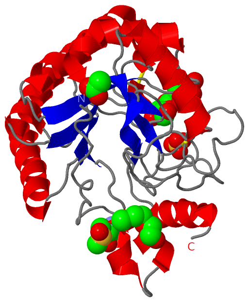
   Legend:   → Mismatch (orange background)
  - → Gap (green background, '-', border residues have a numbering label)
    → Modified Residue (blue background, lower-case, 'x' indicates undefined single-letter code, labelled with number + name)
  x → Chemical Group (purple background, 'x', labelled with number + name, e.g. ACE or NH2)
  extra numbering lines below/above indicate numbering irregularities and modified residue names etc., number ends below/above '|'

 Classification and Annotation

(-) SCOP Domains  (1, 1)

Asymmetric/Biological Unit

(-) CATH Domains  (1, 1)

Asymmetric/Biological Unit
(-)
Class: Alpha Beta (26913)

(-) Pfam Domains  (1, 1)

Asymmetric/Biological Unit

(-) Gene Ontology  (11, 11)

Asymmetric/Biological Unit(hide GO term definitions)
Chain A   (Q79G13_MYCTU | Q79G13)
molecular function
    GO:0008810    cellulase activity    Catalysis of the endohydrolysis of (1->4)-beta-D-glucosidic linkages in cellulose, lichenin and cereal beta-D-glucans.
    GO:0016787    hydrolase activity    Catalysis of the hydrolysis of various bonds, e.g. C-O, C-N, C-C, phosphoric anhydride bonds, etc. Hydrolase is the systematic name for any enzyme of EC class 3.
    GO:0016798    hydrolase activity, acting on glycosyl bonds    Catalysis of the hydrolysis of any glycosyl bond.
    GO:0004553    hydrolase activity, hydrolyzing O-glycosyl compounds    Catalysis of the hydrolysis of any O-glycosyl bond.
biological process
    GO:0005975    carbohydrate metabolic process    The chemical reactions and pathways involving carbohydrates, any of a group of organic compounds based of the general formula Cx(H2O)y. Includes the formation of carbohydrate derivatives by the addition of a carbohydrate residue to another molecule.
    GO:0030245    cellulose catabolic process    The chemical reactions and pathways resulting in the breakdown of cellulose, a linear beta1-4 glucan of molecular mass 50-400 kDa with the pyranose units in the -4C1 conformation.
    GO:0008152    metabolic process    The chemical reactions and pathways, including anabolism and catabolism, by which living organisms transform chemical substances. Metabolic processes typically transform small molecules, but also include macromolecular processes such as DNA repair and replication, and protein synthesis and degradation.
    GO:0000272    polysaccharide catabolic process    The chemical reactions and pathways resulting in the breakdown of a polysaccharide, a polymer of many (typically more than 10) monosaccharide residues linked glycosidically.
cellular component
    GO:0005576    extracellular region    The space external to the outermost structure of a cell. For cells without external protective or external encapsulating structures this refers to space outside of the plasma membrane. This term covers the host cell environment outside an intracellular parasite.
    GO:0016021    integral component of membrane    The component of a membrane consisting of the gene products and protein complexes having at least some part of their peptide sequence embedded in the hydrophobic region of the membrane.
    GO:0016020    membrane    A lipid bilayer along with all the proteins and protein complexes embedded in it an attached to it.

 Visualization

(-) Interactive Views

Asymmetric/Biological Unit
  Complete Structure
    Jena3D(integrated viewing of ligand, site, SAP, PROSITE, SCOP information)
    WebMol | AstexViewer[tm]@PDBe
(Java Applets, require no local installation except for Java; loading may be slow)
    STRAP
(Java WebStart application, automatic local installation, requires Java; full application with system access!)
    RasMol
(require local installation)
    Molscript (VRML)
(requires installation of a VRML viewer; select preferred view via VRML and generate a mono or stereo PDF format file)
 
  Ligands, Modified Residues, Ions
    1PG  [ RasMol | Jena3D ]  +environment [ RasMol | Jena3D ]
    ACT  [ RasMol | Jena3D ]  +environment [ RasMol | Jena3D ]
    BGC  [ RasMol | Jena3D ]  +environment [ RasMol | Jena3D ]
    MHO  [ RasMol | Jena3D ]  +environment [ RasMol | Jena3D ]
    SO4  [ RasMol | Jena3D ]  +environment [ RasMol | Jena3D ]
 
  Sites
    AC1  [ RasMol ]  +environment [ RasMol ]
    AC2  [ RasMol ]  +environment [ RasMol ]
    AC3  [ RasMol ]  +environment [ RasMol ]
    AC4  [ RasMol ]  +environment [ RasMol ]
    AC5  [ RasMol ]  +environment [ RasMol ]
 
  Cis Peptide Bonds
    Asn A:112 - Pro A:113   [ RasMol ]  
 

(-) Still Images

Jmol
  protein: cartoon or spacefill or dots and stick; nucleic acid: cartoon and stick; ligands: spacefill; active site: stick
Molscript
  protein, nucleic acid: cartoon; ligands: spacefill; active site: ball and stick

 Databases and Analysis Tools

(-) Databases

Access by PDB/NDB ID
  1up0
    Family and Domain InformationProDom | SYSTERS
    General Structural InformationGlycoscienceDB | MMDB | NDB | OCA | PDB | PDBe | PDBj | PDBsum | PDBWiki | PQS | PROTEOPEDIA
    Orientation in MembranesOPM
    Protein SurfaceSURFACE
    Secondary StructureDSSP (structure derived) | HSSP (homology derived)
    Structural GenomicsGeneCensus
    Structural NeighboursCE | VAST
    Structure ClassificationCATH | Dali | SCOP
    Validation and Original DataBMRB Data View | BMRB Restraints Grid | EDS | PROCHECK | RECOORD | WHAT_CHECK
 
Access by UniProt ID/Accession number
  Q79G13_MYCTU | Q79G13
    Comparative Protein Structure ModelsModBase
    Genomic InformationEnsembl
    Protein-protein InteractionDIP
    Sequence, Family and Domain InformationInterPro | Pfam | SMART | UniProtKB/TrEMBL
 
Access by Enzyme Classificator   (EC Number)
  (no 'Enzyme Classificator' available)
    General Enzyme InformationBRENDA | EC-PDB | Enzyme | IntEnz
    PathwayKEGG | MetaCyc
 
Access by Disease Identifier   (MIM ID)
  (no 'MIM ID' available)
    Disease InformationOMIM
 
Access by GenAge ID
  (no 'GenAge ID' available)
    Age Related InformationGenAge

(-) Analysis Tools

Access by PDB/NDB ID
    Domain InformationXDom
    Interatomic Contacts of Structural UnitsCSU
    Ligand-protein ContactsLPC
    Protein CavitiescastP
    Sequence and Secondary StructurePDBCartoon
    Structure AlignmentSTRAP(Java WebStart application, automatic local installation, requires Java; full application with system access!)
    Structure and Sequence BrowserSTING
 
Access by UniProt ID/Accession number
  Q79G13_MYCTU | Q79G13
    Protein Disorder PredictionDisEMBL | FoldIndex | GLOBPLOT (for more information see DisProt)

 Related Entries

(-) Entries Sharing at Least One Protein Chain (UniProt ID)

UniProtKB/TrEMBL
        Q79G13_MYCTU | Q79G131uoz 1up2 1up3

(-) Related Entries Specified in the PDB File

1uoz STRUCTURE OF THE ENDOGLUCANASE CEL6 FROM MYCOBACTERIUM TUBERCULOSIS IN COMPLEX WITH THIOCELLOPENTAOSE AT 1.1 ANGSTROM
1up2 STRUCTURE OF THE ENDOGLUCANASE CEL6 FROM MYCOBACTERIUM TUBERCULOSIS IN COMPLEX WITH GLUCOSE-ISOFAGOMINE AT 1.9 ANGSTROM
1up3 STRUCTURE OF THE ENDOGLUCANASE CEL6 FROM MYCOBACTERIUM TUBERCULOSIS IN COMPLEX WITH METHYL-CELLOBIOSYL-4-DEOXY-4-THIO-BETA-D- CELLOBIOSIDE AT 1.6 ANGSTROM