|
|
|
|
 Description
|
|
Compounds
|
||||||||||||||||||||||||
Chains, Units
Summary Information (see also Sequences/Alignments below) |
Ligands, Modified Residues, Ions (1, 6)
Asymmetric Unit (1, 6)
|
Sites (6, 6)
Asymmetric Unit (6, 6)
|
SS Bonds (16, 16)
Asymmetric Unit
|
||||||||||||||||||||||||||||||||||||||||||||||||||||||||||||||||||||
Cis Peptide Bonds (0, 0)| (no "Cis Peptide Bond" information available for 1ULM) |
SAPs(SNPs)/Variants (0, 0)| (no "SAP(SNP)/Variant" information available for 1ULM) |
PROSITE Motifs (2, 6)
Asymmetric Unit (2, 6)
|
||||||||||||||||||||||||||||||||||||||||||||||||||||||||||||||||||||||||||||||||||||||||||||||||
Exons (0, 0)| (no "Exon" information available for 1ULM) |
Sequences/Alignments
Asymmetric UnitChain A from PDB Type:PROTEIN Length:82 aligned with LED2_PHYAM | P83790 from UniProtKB/Swiss-Prot Length:82 Alignment length:82 10 20 30 40 50 60 70 80 LED2_PHYAM 1 APECGERASGKRCPNGKCCSQWGYCGTTDNYCGQGCQSQCDYWRCGRDFGGRLCEEDMCCSKYGWCGYSDDHCEDGCQSQCD 82 SCOP domains d1ulma1 A:1-42 Lectin-D d1ulma2 A:43-82 Lectin-D SCOP domains CATH domains 1ulmA01 A:1-40 1ulmA02 A:41-82 CATH domains Pfam domains ---------------------------------------------------------------------------------- Pfam domains SAPs(SNPs) ---------------------------------------------------------------------------------- SAPs(SNPs) PROSITE (1) CHIT_BIND_I_2 PDB: A:1-42 UniProt: 1-42 -CHIT_BIND_I_2 PDB: A:44-82 PROSITE (1) PROSITE (2) ------------CHIT_BIND_I_1 -------------------------------------------------- PROSITE (2) Transcript ---------------------------------------------------------------------------------- Transcript 1ulm A 1 APECGERASGKRCPNGKCCSQWGYCGTTDNYCGQGCQSQCDYWRCGRDFGGRLCEEDMCCSKYGWCGYSDDHCEDGCQSQCD 82 10 20 30 40 50 60 70 80 Chain B from PDB Type:PROTEIN Length:82 aligned with LED2_PHYAM | P83790 from UniProtKB/Swiss-Prot Length:82 Alignment length:82 10 20 30 40 50 60 70 80 LED2_PHYAM 1 APECGERASGKRCPNGKCCSQWGYCGTTDNYCGQGCQSQCDYWRCGRDFGGRLCEEDMCCSKYGWCGYSDDHCEDGCQSQCD 82 SCOP domains d1ulmb1 B:101-142 Lectin-D d1ulmb2 B:143-182 Lectin-D SCOP domains CATH domains 1ulmB01 B:101-140 1ulmB02 B:141-182 CATH domains Pfam domains (1) ------------------------------------------Chitin_bind_1-1ulmB01 B:143-181 - Pfam domains (1) Pfam domains (2) ------------------------------------------Chitin_bind_1-1ulmB02 B:143-181 - Pfam domains (2) Pfam domains (3) ------------------------------------------Chitin_bind_1-1ulmB03 B:143-181 - Pfam domains (3) Pfam domains (4) ------------------------------------------Chitin_bind_1-1ulmB04 B:143-181 - Pfam domains (4) SAPs(SNPs) ---------------------------------------------------------------------------------- SAPs(SNPs) PROSITE (1) CHIT_BIND_I_2 PDB: B:101-142 -CHIT_BIND_I_2 PDB: B:144-182 PROSITE (1) PROSITE (2) ------------CHIT_BIND_I_1 -------------------------------------------------- PROSITE (2) Transcript ---------------------------------------------------------------------------------- Transcript 1ulm B 101 APECGERASGKRCPNGKCCSQWGYCGTTDNYCGQGCQSQCDYWRCGRDFGGRLCEEDMCCSKYGWCGYSDDHCEDGCQSQCD 182 110 120 130 140 150 160 170 180
|
||||||||||||||||||||
SCOP Domains (1, 4)
Asymmetric Unit
 |
CATH Domains (1, 4)
Asymmetric Unit
|
Pfam Domains (1, 4)
Asymmetric Unit
|
Gene Ontology (4, 4)|
Asymmetric Unit(hide GO term definitions) Chain A,B (LED2_PHYAM | P83790)
|
||||||||||||||||||||||||||||||||||||
Interactive Views
|
||||||||||||||||||||||||||||||||||||||||||||||||||||||||||||||||||||||||||||||||||||||||||||||||||||||||||||||||||||||||||||||||||||||||||||||||||||||||||||||||||||||||||||||||
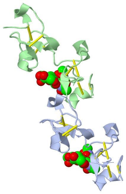
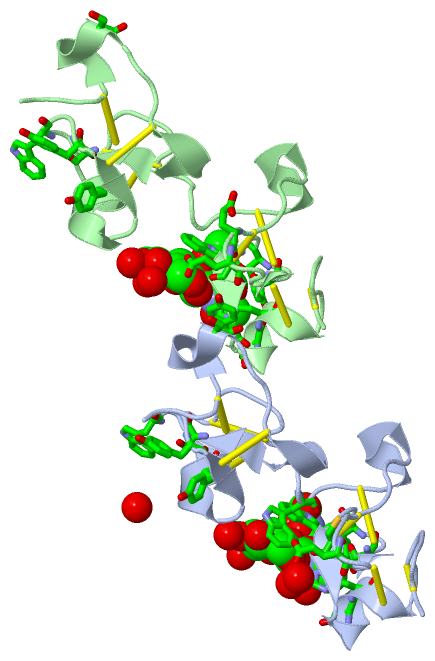
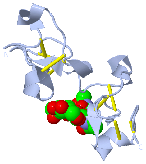
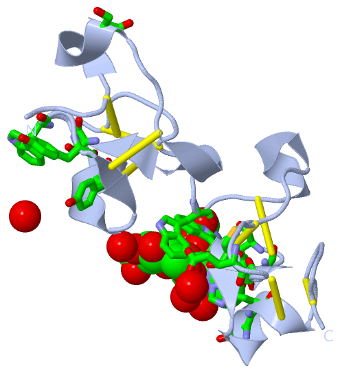
Still Images
|
||||||||||||||||
Databases
|
||||||||||||||||||||||||||||||||||||||||||||||||||||||||||||||||||||||||||||||||||||||||||||||||||||||||||||||||||||||||||||||||||||||||||||||||||||||||||||||||
Analysis Tools
|
|||||||||||||||||||||||||||||||||||||||||||||||||||||||||||||
Entries Sharing at Least One Protein Chain (UniProt ID)
Related Entries Specified in the PDB File
|
|