|
|
|
|
 Description
|
|
Compounds
|
||||||||||||||||||||||||||||||||||||||||||||||||||||||||||||||||||||||||||||||||||||||||||||||||||||||||||||||||||||||||||||||
Chains, Units
Summary Information (see also Sequences/Alignments below) |
Ligands, Modified Residues, Ions (2, 2)| NMR Structure (2, 2) NMR Structure * (1, 1) |
Sites (2, 2)
NMR Structure (2, 2)
|
SS Bonds (0, 0)| (no "SS Bond" information available for 1TU2) |
Cis Peptide Bonds (3, 30)
NMR Structure
|
||||||||||||||||||||
SAPs(SNPs)/Variants (0, 0)| (no "SAP(SNP)/Variant" information available for 1TU2) |
PROSITE Motifs (1, 1)
NMR Structure (1, 1)
|
||||||||||||||||||||||||||||||||||||||||||||||||
Exons (0, 0)| (no "Exon" information available for 1TU2) |
Sequences/Alignments
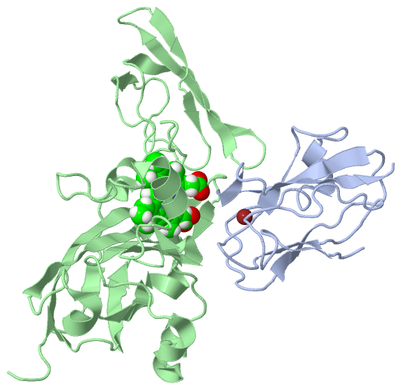
NMR StructureChain A from PDB Type:PROTEIN Length:105 aligned with PLAS_NOSS1 | P46444 from UniProtKB/Swiss-Prot Length:139 Alignment length:105 44 54 64 74 84 94 104 114 124 134 PLAS_NOSS1 35 ETYTVKLGSDKGLLVFEPAKLTIKPGDTVEFLNNKVPPHNVVFDAALNPAKSADLAKSLSHKQLLMSPGQSTSTTFPADAPAGEYTFYCEPHRGAGMVGKITVAG 139 SCOP domains d1tu2a1 A:1-105 Plastocyanin SCOP domains CATH domains 1tu2A00 A:1-105 Cupredoxins - blue copper proteins CATH domains Pfam domains --Copper-bind-1tu2A01 A:3-104 - Pfam domains SAPs(SNPs) --------------------------------------------------------------------------------------------------------- SAPs(SNPs) PROSITE ---------------------------------------------------------------------------------COPPER_BLUE -------- PROSITE Transcript --------------------------------------------------------------------------------------------------------- Transcript 1tu2 A 1 ETYTVKLGSDKGLLVFEPAKLTIKPGDTVEFLNNKVPPHNVVFDAALNPAKSADLAKSLSHKQLLMSPGQSTSTTFPADAPAGEYTFYCEPHRGAGMVGKITVAG 105 10 20 30 40 50 60 70 80 90 100 Chain B from PDB Type:PROTEIN Length:254 aligned with CYF_NOSS1 | Q93SW9 from UniProtKB/Swiss-Prot Length:333 Alignment length:254 54 64 74 84 94 104 114 124 134 144 154 164 174 184 194 204 214 224 234 244 254 264 274 284 294 CYF_NOSS1 45 YPFWAQQTYPETPREPTGRIVCANCHLAAKPTEVEVPQSVLPDTVFKAVVKIPYDTSVQQVGADGSKVGLNVGAVLMLPEGFKIAPEDRIPEELKEEIGDVYFQPYGEDKDNIVIVGPLPGEQYQEIVFPVLSPNPANDKNIHFGKYSVHVGGNRGRGQVYPTGEKSNNNLYSAAATGTISKIAKQEGEDGSVKYLVDIKTESGEVVSDTIPAGPELIVSEGQAVTAGDALTNNPNVGGFGQLDAEIVLQDANR 298 SCOP domains d1tu2b1 B:1-169,B:236-254 Cytochrome f, large domain d1tu2b2 B:170-235 Cytochrome f, small domain d1tu2b1 SCOP domains CATH domains 1tu2B01 B:1-171,B:234-252 [code=2.60.40.830, no name defined] 1tu2B02 B:172-233 [code=2.40.50.100, no name defined] 1tu2B01 -- CATH domains Pfam domains -----------------------------------------------------------------------------------------------------------------------------------------------------------------------Apocytochr_F_C-1tu2B01 B:168-254 Pfam domains SAPs(SNPs) -------------------------------------------------------------------------------------------------------------------------------------------------------------------------------------------------------------------------------------------------------------- SAPs(SNPs) PROSITE -------------------------------------------------------------------------------------------------------------------------------------------------------------------------------------------------------------------------------------------------------------- PROSITE Transcript -------------------------------------------------------------------------------------------------------------------------------------------------------------------------------------------------------------------------------------------------------------- Transcript 1tu2 B 1 YPFWAQQTYPETPREPTGRIVCANCHLAAKPTEVEVPQSVLPDTVFKAVVKIPYDTSVQQVGADGSKVGLNVGAVLMLPEGFKIAPEDRIPEELKEEIGDVYFQPYGEDKDNIVIVGPLPGEQYQEIVFPVLSPNPANDKNIHFGKYSVHVGGNRGRGQVYPTGEKSNNNLYSAAATGTISKIAKQEGEDGSVKYLVDIKTESGEVVSDTIPAGPELIVSEGQAVTAGDALTNNPNVGGFGQLDAEIVLQDANR 254 10 20 30 40 50 60 70 80 90 100 110 120 130 140 150 160 170 180 190 200 210 220 230 240 250
|
||||||||||||||||||||
SCOP Domains (3, 3)
NMR Structure

|
CATH Domains (3, 3)
NMR Structure
|
Pfam Domains (2, 2)
NMR Structure
|
Gene Ontology (12, 18)|
NMR Structure(hide GO term definitions) Chain A (PLAS_NOSS1 | P46444)
Chain B (CYF_NOSS1 | Q93SW9)
|
||||||||||||||||||||||||||||||||||||||||||||||||||||||||||||||||||||||||||||||||||||||||||||||||||||||||||||||||||||||||||||||||||||||||||||||||
Interactive Views
|
|||||||||||||||||||||||||||||||||||||||||||||||||||||||||||||||||||||||||||||||||||||||||||||||||||||||||||||||||||||||||||||||||||||||||||||||||||
Still Images
|
||||||||||||||||
Databases
|
||||||||||||||||||||||||||||||||||||||||||||||||||||||||||||||||||||||||||||||||||||||||||||||||||||||||||||||||||||||||||||||||||||||||||||||||||||||||||||||||||||||||||||||||||||||||||
Analysis Tools
|
||||||||||||||||||||||||||||||||||||||||||||||||||||||||||||||||||||||||
Entries Sharing at Least One Protein Chain (UniProt ID)
Related Entries Specified in the PDB File
|
|