|
|
|
|
 Description
|
|
Compounds
|
||||||||||||||||||||||||||||
Chains, Units
Summary Information (see also Sequences/Alignments below) |
Ligands, Modified Residues, Ions (4, 8)| Asymmetric/Biological Unit (4, 8) |
Sites (14, 14)
Asymmetric Unit (14, 14)
|
SS Bonds (1, 1)
Asymmetric/Biological Unit
|
||||||||
Cis Peptide Bonds (0, 0)| (no "Cis Peptide Bond" information available for 1SLA) |
SAPs(SNPs)/Variants (0, 0)| (no "SAP(SNP)/Variant" information available for 1SLA) |
PROSITE Motifs (1, 2)
Asymmetric/Biological Unit (1, 2)
|
||||||||||||||||||||||||
Exons (2, 4)
Asymmetric/Biological Unit (2, 4)
|
||||||||||||||||||||||||||||||||||||||||||||||||
Sequences/Alignments
Asymmetric/Biological UnitChain A from PDB Type:PROTEIN Length:134 aligned with LEG1_BOVIN | P11116 from UniProtKB/Swiss-Prot Length:135 Alignment length:134 11 21 31 41 51 61 71 81 91 101 111 121 131 LEG1_BOVIN 2 ACGLVASNLNLKPGECLRVRGEVAADAKSFLLNLGKDDNNLCLHFNPRFNAHGDVNTIVCNSKDAGAWGAEQRESAFPFQPGSVVEVCISFNQTDLTIKLPDGYEFKFPNRLNLEAINYLSAGGDFKIKCVAFE 135 SCOP domains d1slaa_ A: Galectin-1 SCOP domains CATH domains 1slaA00 A:1-134 [code=2.60.120.200, no name defined] CATH domains Pfam domains -------------------------------------------------------------------------------------------------------------------------------------- Pfam domains SAPs(SNPs) -------------------------------------------------------------------------------------------------------------------------------------- SAPs(SNPs) PROSITE --GALECTIN PDB: A:3-134 UniProt: 4-135 PROSITE Transcript 1 Exon 1.1 PDB: A:1-57 UniProt: 1-58 [INCOMPLETE] Exon 1.2 PDB: A:58-105 UniProt: 59-106 ----------------------------- Transcript 1 1sla A 1 ACGLVASNLNLKPGECLRVRGEVAADAKSFLLNLGKDDNNLCLHFNPRFNAHGDVNTIVCNSKDAGAWGAEQRESAFPFQPGSVVEVCISFNQTDLTIKLPDGYEFKFPNRLNLEAINYLSAGGDFKIKCVAFE 134 10 20 30 40 50 60 70 80 90 100 110 120 130 Chain B from PDB Type:PROTEIN Length:134 aligned with LEG1_BOVIN | P11116 from UniProtKB/Swiss-Prot Length:135 Alignment length:134 11 21 31 41 51 61 71 81 91 101 111 121 131 LEG1_BOVIN 2 ACGLVASNLNLKPGECLRVRGEVAADAKSFLLNLGKDDNNLCLHFNPRFNAHGDVNTIVCNSKDAGAWGAEQRESAFPFQPGSVVEVCISFNQTDLTIKLPDGYEFKFPNRLNLEAINYLSAGGDFKIKCVAFE 135 SCOP domains d1slab_ B: Galectin-1 SCOP domains CATH domains 1slaB00 B:1-134 [code=2.60.120.200, no name defined] CATH domains Pfam domains (1) --Gal-bind_lectin-1slaB01 B:3-133 - Pfam domains (1) Pfam domains (2) --Gal-bind_lectin-1slaB02 B:3-133 - Pfam domains (2) SAPs(SNPs) -------------------------------------------------------------------------------------------------------------------------------------- SAPs(SNPs) PROSITE --GALECTIN PDB: B:3-134 UniProt: 4-135 PROSITE Transcript 1 Exon 1.1 PDB: B:1-57 UniProt: 1-58 [INCOMPLETE] Exon 1.2 PDB: B:58-105 UniProt: 59-106 ----------------------------- Transcript 1 1sla B 1 ACGLVASNLNLKPGECLRVRGEVAADAKSFLLNLGKDDNNLCLHFNPRFNAHGDVNTIVCNSKDAGAWGAEQRESAFPFQPGSVVEVCISFNQTDLTIKLPDGYEFKFPNRLNLEAINYLSAGGDFKIKCVAFE 134 10 20 30 40 50 60 70 80 90 100 110 120 130
|
||||||||||||||||||||
SCOP Domains (1, 2)
Asymmetric/Biological Unit
 |
CATH Domains (1, 2)
Asymmetric/Biological Unit
|
Pfam Domains (1, 2)
Asymmetric/Biological Unit
|
Gene Ontology (15, 15)|
Asymmetric/Biological Unit(hide GO term definitions) Chain A,B (LEG1_BOVIN | P11116)
|
||||||||||||||||||||||||||||||||||||||||||||||||||||||||||||||||||||||||||||||||||||||||||||||||||||||||||||
Interactive Views
|
||||||||||||||||||||||||||||||||||||||||||||||||||||||||||||||||||||||||||||||||||||||||||||||||||||||||||||||||||||||||||||||||||||||||||||||||||||||||||||||||||||||||||||||||||||||||||||||||||||||||||||||||||||||||||||||||||||||
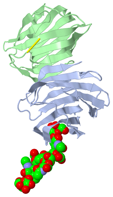
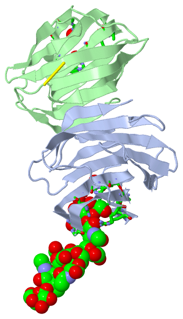
Still Images
|
||||||||||||||||
Databases
|
||||||||||||||||||||||||||||||||||||||||||||||||||||||||||||||||||||||||||||||||||||||||||||||||||||||||||||||||||||||||||||||||||||||||||||||||||||||||||||||||
Analysis Tools
|
|||||||||||||||||||||||||||||||||||||||||||||||||||||||||||||
Entries Sharing at Least One Protein Chain (UniProt ID)
Related Entries Specified in the PDB File
|
|