Show PDB file:   
         Plain Text   HTML   (compressed file size)
QuickSearch:   
by PDB,NDB,UniProt,PROSITE Code or Search Term(s)  
(-)Biol.Unit 1 - manually
(-)Asym.Unit - manually
(-)Asymmetric Unit
(-)Biological Unit 1
collapse expand < >
Image Biol.Unit 1 - manually
Biol.Unit 1 - manually  (Jmol Viewer)
Image Asym.Unit - manually
Asym.Unit - manually  (Jmol Viewer)
Image Asymmetric Unit
Asymmetric Unit  (Jmol Viewer)
Image Biological Unit 1
Biological Unit 1  (Jmol Viewer)

(-) Description

Title :  CRYSTAL STRUCTURE OF DCOH2, A PARALOG OF DCOH, THE DIMERIZATION COFACTOR OF HNF-1
 
Authors :  R. B. Rose, K. E. Pullen, J. H. Bayle, G. R. Crabtree, T. Alber
Date :  10 Dec 03  (Deposition) - 12 Oct 04  (Release) - 24 Feb 09  (Revision)
Method :  X-RAY DIFFRACTION
Resolution :  1.60
Chains :  Asym. Unit :  A,B
Biol. Unit 1:  A,B  (2x)
Keywords :  Alpha And Beta Structure, Lyase (Keyword Search: [Gene Ontology, PubMed, Web (Google))
 
Reference :  R. B. Rose, K. E. Pullen, J. H. Bayle, G. R. Crabtree, T. Alber
Biochemical And Structural Basis For Partially Redundant Enzymatic And Transcriptional Functions Of Dcoh And Dcoh2
Biochemistry V. 43 7345 2004
PubMed-ID: 15182178  |  Reference-DOI: 10.1021/BI049620T
(for further references see the PDB file header)

(-) Compounds

Molecule 1 - DCOH-LIKE PROTEIN DCOHM
    ChainsA, B
    EC Number4.2.1.96
    EngineeredYES
    Expression SystemESCHERICHIA COLI BL21(DE3)
    Expression System PlasmidPGEX2T
    Expression System StrainBL21(DE3)
    Expression System Taxid469008
    Expression System Vector TypePLASMID
    GeneDCOHM
    OrganLIVER
    Organism CommonHOUSE MOUSE
    Organism ScientificMUS MUSCULUS
    Organism Taxid10090

 Structural Features

(-) Chains, Units

  12
Asymmetric Unit AB
Biological Unit 1 (2x)AB

Summary Information (see also Sequences/Alignments below)

(-) Ligands, Modified Residues, Ions  (0, 0)

(no "Ligand,Modified Residues,Ions" information available for 1RU0)

(-) Sites  (0, 0)

(no "Site" information available for 1RU0)

(-) SS Bonds  (0, 0)

(no "SS Bond" information available for 1RU0)

(-) Cis Peptide Bonds  (0, 0)

(no "Cis Peptide Bond" information available for 1RU0)

 Sequence-Structure Mapping

(-) SAPs(SNPs)/Variants  (0, 0)

(no "SAP(SNP)/Variant" information available for 1RU0)

(-) PROSITE Motifs  (0, 0)

(no "PROSITE Motif" information available for 1RU0)

(-) Exons   (0, 0)

(no "Exon" information available for 1RU0)

(-) Sequences/Alignments

Asymmetric Unit
   Reformat: Number of residues per line =  ('0' or empty: single-line sequence representation)
  Number of residues per labelling interval =   
  UniProt sequence: complete  aligned part    
   Show mapping: SCOP domains CATH domains Pfam domains Secondary structure (by author)
SAPs(SNPs) PROSITE motifs Exons
(details for a mapped element are shown in a popup box when the mouse pointer rests over it)
Chain A from PDB  Type:PROTEIN  Length:100
 aligned with PHS2_MOUSE | Q9CZL5 from UniProtKB/Swiss-Prot  Length:136

    Alignment length:100
                                    46        56        66        76        86        96       106       116       126       136
           PHS2_MOUSE    37 DAQWLTAEERDQLIPGLKAAGWSELSERDAIYKEFSFKNFNQAFGFMSRVALQAEKMNHHPEWFNVYNKVQITLTSHDCGGLTKRDVKLAQFIEKAAASL 136
               SCOP domains d1ru0a_ A: DcoH-like protein DCoH2                                                                   SCOP domains
               CATH domains 1ru0A00 A:4-103  [code=3.30.1360.20, no name defined]                                                CATH domains
               Pfam domains ---------------------------------------------------------------------------------------------------- Pfam domains
         Sec.struct. author .....hhhhhhhhhhhhhhh..ee......eeeeee..hhhhhhhhhhhhhhhhhhhh...eeeee..eeeeee........hhhhhhhhhhhhhhhh.. Sec.struct. author
                 SAPs(SNPs) ---------------------------------------------------------------------------------------------------- SAPs(SNPs)
                    PROSITE ---------------------------------------------------------------------------------------------------- PROSITE
                 Transcript ---------------------------------------------------------------------------------------------------- Transcript
                 1ru0 A   4 DAQWLTAEERDQLIPGLKAAGWSELSERDAIYKEFSFKNFNQAFGFMSRVALQAEKMNHHPEWFNVYNKVQITLTSHDCGGLTKRDVKLAQFIEKAAASL 103
                                    13        23        33        43        53        63        73        83        93       103

Chain B from PDB  Type:PROTEIN  Length:98
 aligned with PHS2_MOUSE | Q9CZL5 from UniProtKB/Swiss-Prot  Length:136

    Alignment length:98
                                    46        56        66        76        86        96       106       116       126        
           PHS2_MOUSE    37 DAQWLTAEERDQLIPGLKAAGWSELSERDAIYKEFSFKNFNQAFGFMSRVALQAEKMNHHPEWFNVYNKVQITLTSHDCGGLTKRDVKLAQFIEKAAA 134
               SCOP domains d1ru0b_ B: DcoH-like protein DCoH2                                                                 SCOP domains
               CATH domains 1ru0B00 B:4-101  [code=3.30.1360.20, no name defined]                                              CATH domains
           Pfam domains (1) Pterin_4a-1ru0B01 B:4-101                                                                          Pfam domains (1)
           Pfam domains (2) Pterin_4a-1ru0B02 B:4-101                                                                          Pfam domains (2)
         Sec.struct. author .....hhhhhhhhhhhhhhh..ee......eeeeee..hhhhhhhhhhhhhhhhhhhh...eeeee..eeeeee........hhhhhhhhhhhhhhhh Sec.struct. author
                 SAPs(SNPs) -------------------------------------------------------------------------------------------------- SAPs(SNPs)
                    PROSITE -------------------------------------------------------------------------------------------------- PROSITE
                 Transcript -------------------------------------------------------------------------------------------------- Transcript
                 1ru0 B   4 DAQWLTAEERDQLIPGLKAAGWSELSERDAIYKEFSFKNFNQAFGFMSRVALQAEKMNHHPEWFNVYNKVQITLTSHDCGGLTKRDVKLAQFIEKAAA 101
                                    13        23        33        43        53        63        73        83        93        

   Legend:   → Mismatch (orange background)
  - → Gap (green background, '-', border residues have a numbering label)
    → Modified Residue (blue background, lower-case, 'x' indicates undefined single-letter code, labelled with number + name)
  x → Chemical Group (purple background, 'x', labelled with number + name, e.g. ACE or NH2)
  extra numbering lines below/above indicate numbering irregularities and modified residue names etc., number ends below/above '|'

 Classification and Annotation

(-) SCOP Domains  (1, 2)

Asymmetric Unit

(-) CATH Domains  (1, 2)

Asymmetric Unit
(-)
Class: Alpha Beta (26913)

(-) Pfam Domains  (1, 2)

Asymmetric Unit

(-) Gene Ontology  (11, 11)

Asymmetric Unit(hide GO term definitions)
Chain A,B   (PHS2_MOUSE | Q9CZL5)
molecular function
    GO:0008124    4-alpha-hydroxytetrahydrobiopterin dehydratase activity    Catalysis of the reaction: (6R)-6-(L-erythro-1,2-dihydroxypropyl)-5,6,7,8-tetrahydro-4a-hydroxypterin = (6R)-6-(L-erythro-1,2-dihydroxypropyl)-7,8-dihydro-6H-pterin + H(2)O.
    GO:0016829    lyase activity    Catalysis of the cleavage of C-C, C-O, C-N and other bonds by other means than by hydrolysis or oxidation, or conversely adding a group to a double bond. They differ from other enzymes in that two substrates are involved in one reaction direction, but only one in the other direction. When acting on the single substrate, a molecule is eliminated and this generates either a new double bond or a new ring.
    GO:0004505    phenylalanine 4-monooxygenase activity    Catalysis of the reaction: L-phenylalanine + tetrahydrobiopterin + O2 = L-tyrosine + 4-alpha-hydroxytetrahydrobiopterin.
    GO:0005515    protein binding    Interacting selectively and non-covalently with any protein or protein complex (a complex of two or more proteins that may include other nonprotein molecules).
biological process
    GO:0055114    oxidation-reduction process    A metabolic process that results in the removal or addition of one or more electrons to or from a substance, with or without the concomitant removal or addition of a proton or protons.
    GO:0045893    positive regulation of transcription, DNA-templated    Any process that activates or increases the frequency, rate or extent of cellular DNA-templated transcription.
    GO:0051291    protein heterooligomerization    The process of creating protein oligomers, compounds composed of a small number, usually between three and ten, of component monomers that are not all identical. Oligomers may be formed by the polymerization of a number of monomers or the depolymerization of a large protein polymer.
    GO:0051289    protein homotetramerization    The formation of a protein homotetramer, a macromolecular structure consisting of four noncovalently associated identical subunits.
    GO:0006729    tetrahydrobiopterin biosynthetic process    The chemical reactions and pathways resulting in the formation of tetrahydrobiopterin, the reduced form of biopterin (2-amino-4-hydroxy-6-(1,2-dihydroxypropyl)-pteridine). It functions as a hydroxylation coenzyme, e.g. in the conversion of phenylalanine to tyrosine.
cellular component
    GO:0005739    mitochondrion    A semiautonomous, self replicating organelle that occurs in varying numbers, shapes, and sizes in the cytoplasm of virtually all eukaryotic cells. It is notably the site of tissue respiration.
    GO:0005634    nucleus    A membrane-bounded organelle of eukaryotic cells in which chromosomes are housed and replicated. In most cells, the nucleus contains all of the cell's chromosomes except the organellar chromosomes, and is the site of RNA synthesis and processing. In some species, or in specialized cell types, RNA metabolism or DNA replication may be absent.
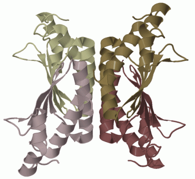
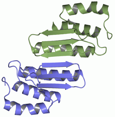
 Visualization

(-) Interactive Views

Asymmetric Unit
  Complete Structure
    Jena3D(integrated viewing of ligand, site, SAP, PROSITE, SCOP information)
    WebMol | AstexViewer[tm]@PDBe
(Java Applets, require no local installation except for Java; loading may be slow)
    STRAP
(Java WebStart application, automatic local installation, requires Java; full application with system access!)
    RasMol
(require local installation)
    Molscript (VRML)
(requires installation of a VRML viewer; select preferred view via VRML and generate a mono or stereo PDF format file)
 
  Ligands, Modified Residues, Ions
(no "Ligands, Modified Residues, Ions" information available for 1ru0)
 
  Sites
(no "Sites" information available for 1ru0)
 
  Cis Peptide Bonds
(no "Cis Peptide Bonds" information available for 1ru0)
 
Biological Unit
  Complete Structure
    Biological Unit 1  [ Jena3D ]

(-) Still Images

Jmol
  protein: cartoon or spacefill or dots and stick; nucleic acid: cartoon and stick; ligands: spacefill; active site: stick
Molscript
  protein, nucleic acid: cartoon; ligands: spacefill; active site: ball and stick

 Databases and Analysis Tools

(-) Databases

Access by PDB/NDB ID
  1ru0
    Family and Domain InformationProDom | SYSTERS
    General Structural InformationGlycoscienceDB | MMDB | NDB | OCA | PDB | PDBe | PDBj | PDBsum | PDBWiki | PQS | PROTEOPEDIA
    Orientation in MembranesOPM
    Protein SurfaceSURFACE
    Secondary StructureDSSP (structure derived) | HSSP (homology derived)
    Structural GenomicsGeneCensus
    Structural NeighboursCE | VAST
    Structure ClassificationCATH | Dali | SCOP
    Validation and Original DataBMRB Data View | BMRB Restraints Grid | EDS | PROCHECK | RECOORD | WHAT_CHECK
 
Access by UniProt ID/Accession number
  PHS2_MOUSE | Q9CZL5
    Comparative Protein Structure ModelsModBase
    Genomic InformationEnsembl
    Protein-protein InteractionDIP
    Sequence, Family and Domain InformationInterPro | Pfam | SMART | UniProtKB/SwissProt
 
Access by Enzyme Classificator   (EC Number)
  4.2.1.96
    General Enzyme InformationBRENDA | EC-PDB | Enzyme | IntEnz
    PathwayKEGG | MetaCyc
 
Access by Disease Identifier   (MIM ID)
  (no 'MIM ID' available)
    Disease InformationOMIM
 
Access by GenAge ID
  (no 'GenAge ID' available)
    Age Related InformationGenAge

(-) Analysis Tools

Access by PDB/NDB ID
    Domain InformationXDom
    Interatomic Contacts of Structural UnitsCSU
    Ligand-protein ContactsLPC
    Protein CavitiescastP
    Sequence and Secondary StructurePDBCartoon
    Structure AlignmentSTRAP(Java WebStart application, automatic local installation, requires Java; full application with system access!)
    Structure and Sequence BrowserSTING
 
Access by UniProt ID/Accession number
  PHS2_MOUSE | Q9CZL5
    Protein Disorder PredictionDisEMBL | FoldIndex | GLOBPLOT (for more information see DisProt)

 Related Entries

(-) Entries Sharing at Least One Protein Chain (UniProt ID)

UniProtKB/Swiss-Prot
        PHS2_MOUSE | Q9CZL54wil

(-) Related Entries Specified in the PDB File

(no "Related Entries Specified in the PDB File" available for 1RU0)