|
|
|
|
 Description
|
|
Compounds
|
||||||||||||||||||||||||||||||||||||||||||||||||||||
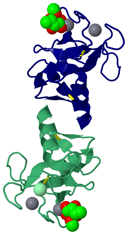
Chains, Units
Summary Information (see also Sequences/Alignments below) |
Ligands, Modified Residues, Ions (3, 7)| Asymmetric/Biological Unit (3, 7) |
Sites (11, 11)
Asymmetric Unit (11, 11)
|
SS Bonds (4, 4)
Asymmetric/Biological Unit
|
||||||||||||||||||||
Cis Peptide Bonds (2, 2)
Asymmetric/Biological Unit
|
||||||||||||
SAPs(SNPs)/Variants (0, 0)| (no "SAP(SNP)/Variant" information available for 1RDI) |
PROSITE Motifs (2, 4)
Asymmetric/Biological Unit (2, 4)
|
||||||||||||||||||||||||||||||||
Exons (0, 0)| (no "Exon" information available for 1RDI) |
Sequences/Alignments
Asymmetric/Biological UnitChain 1 from PDB Type:PROTEIN Length:111 aligned with MBL2_RAT | P08661 from UniProtKB/Swiss-Prot Length:244 Alignment length:111 142 152 162 172 182 192 202 212 222 232 242 MBL2_RAT 133 KYFMSSVRRMPLNRAKALCSELQGTVATPRNAEENRAIQNVAKDVAFLGITDQRTENVFEDLTGNRVRYTNWNEGEPNNVGSGENCVVLLTNGKWNDVPCSDSFLVVCEFS 243 SCOP domains d1rdi1_ 1: Mannose-binding protein A, C-lectin domain SCOP domains CATH domains 1rdi100 1:115-225 Mannose-Binding Protein A, subunit A CATH domains Pfam domains --------------------------------------------------------------------------------------------------------------- Pfam domains SAPs(SNPs) --------------------------------------------------------------------------------------------------------------- SAPs(SNPs) PROSITE (1) -C_TYPE_LECTIN_2 PDB: 1:116-223 UniProt: 134-241 -- PROSITE (1) PROSITE (2) -------------------------------------------------------------------------------------C_TYPE_LECTIN_1 --- PROSITE (2) Transcript --------------------------------------------------------------------------------------------------------------- Transcript 1rdi 1 115 KYFMSSVRRMPLNRAKALCSELQGTVATPRNAEENRAIQNVAKDVAFLGITDQRTENVFEDLTGNRVRYTNWNEGEPNNVGSGENCVVLLTNGKWNDVPCSDSFLVVCEFS 225 124 134 144 154 164 174 184 194 204 214 224 Chain 2 from PDB Type:PROTEIN Length:112 aligned with MBL2_RAT | P08661 from UniProtKB/Swiss-Prot Length:244 Alignment length:112 141 151 161 171 181 191 201 211 221 231 241 MBL2_RAT 132 KKYFMSSVRRMPLNRAKALCSELQGTVATPRNAEENRAIQNVAKDVAFLGITDQRTENVFEDLTGNRVRYTNWNEGEPNNVGSGENCVVLLTNGKWNDVPCSDSFLVVCEFS 243 SCOP domains d1rdi2_ 2: Mannose-binding protein A, C-lectin domain SCOP domains CATH domains 1rdi200 2:114-225 Mannose-Binding Protein A, subunit A CATH domains Pfam domains (1) --------Lectin_C-1rdi201 2:122-224 - Pfam domains (1) Pfam domains (2) --------Lectin_C-1rdi202 2:122-224 - Pfam domains (2) SAPs(SNPs) ---------------------------------------------------------------------------------------------------------------- SAPs(SNPs) PROSITE (1) --C_TYPE_LECTIN_2 PDB: 2:116-223 UniProt: 134-241 -- PROSITE (1) PROSITE (2) --------------------------------------------------------------------------------------C_TYPE_LECTIN_1 --- PROSITE (2) Transcript ---------------------------------------------------------------------------------------------------------------- Transcript 1rdi 2 114 KKYFMSSVRRMPLNRAKALCSELQGTVATPRNAEENRAIQNVAKDVAFLGITDQRTENVFEDLTGNRVRYTNWNEGEPNNVGSGENCVVLLTNGKWNDVPCSDSFLVVCEFS 225 123 133 143 153 163 173 183 193 203 213 223
|
||||||||||||||||||||
SCOP Domains (1, 2)
Asymmetric/Biological Unit
 |
CATH Domains (1, 2)
Asymmetric/Biological Unit
|
Pfam Domains (1, 2)
Asymmetric/Biological Unit
|
Gene Ontology (19, 19)|
Asymmetric/Biological Unit(hide GO term definitions) Chain 1,2 (MBL2_RAT | P08661)
|
||||||||||||||||||||||||||||||||||||||||||||||||||||||||||||||||||||||||||||||||||||||||||||||||||||||||||||||||||||||||||||||||||||
Interactive Views
|
||||||||||||||||||||||||||||||||||||||||||||||||||||||||||||||||||||||||||||||||||||||||||||||||||||||||||||||||||||||||||||||||||||||||||||||||||||||||||||||||||||||||||||||||||||||||||||||||||||||||||||||||||
Still Images
|
||||||||||||||||
Databases
|
||||||||||||||||||||||||||||||||||||||||||||||||||||||||||||||||||||||||||||||||||||||||||||||||||||||||||||||||||||||||||||||||||||||||||||||||||||||||||||||||
Analysis Tools
|
|||||||||||||||||||||||||||||||||||||||||||||||||||||||||||||
Entries Sharing at Least One Protein Chain (UniProt ID)
Related Entries Specified in the PDB File
|
|