|
|
|
|
 Description
|
|
Compounds
|
||||||||||||||||||||||||||||||||||||||||||||||
Chains, Units
Summary Information (see also Sequences/Alignments below) |
Ligands, Modified Residues, Ions (1, 4)
Asymmetric/Biological Unit (1, 4)
|
Sites (4, 4)
Asymmetric Unit (4, 4)
|
SS Bonds (3, 3)
Asymmetric/Biological Unit
|
||||||||||||||||
Cis Peptide Bonds (3, 3)
Asymmetric/Biological Unit
|
||||||||||||||||
SAPs(SNPs)/Variants (0, 0)| (no "SAP(SNP)/Variant" information available for 1R0R) |
PROSITE Motifs (4, 4)
Asymmetric/Biological Unit (4, 4)
|
||||||||||||||||||||||||||||||||||||||||||||||||
Exons (0, 0)| (no "Exon" information available for 1R0R) |
Sequences/Alignments
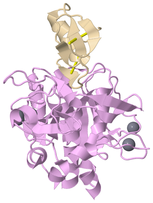
Asymmetric/Biological UnitChain E from PDB Type:PROTEIN Length:274 aligned with SUBT_BACLI | P00780 from UniProtKB/Swiss-Prot Length:379 Alignment length:274 115 125 135 145 155 165 175 185 195 205 215 225 235 245 255 265 275 285 295 305 315 325 335 345 355 365 375 SUBT_BACLI 106 AQTVPYGIPLIKADKVQAQGFKGANVKVAVLDTGIQASHPDLNVVGGASFVAGEAYNTDGNGHGTHVAGTVAALDNTTGVLGVAPSVSLYAVKVLNSSGSGTYSGIVSGIEWATTNGMDVINMSLGGPSGSTAMKQAVDNAYARGVVVVAAAGNSGSSGNTNTIGYPAKYDSVIAVGAVDSNSNRASFSSVGAELEVMAPGAGVYSTYPTSTYATLNGTSMASPHVAGAAALILSKHPNLSASQVRNRLSSTATYLGSSFYYGKGLINVEAAAQ 379 SCOP domains d1r0re_ E: Subtilisin SCOP domains CATH domains 1r0rE00 E:1-275 [code=3.40.50.200, no name defined] CATH domains Pfam domains --------------------------Peptidase_S8-1r0rE01 E:27-275 Pfam domains SAPs(SNPs) ---------------------------------------------------------------------------------------------------------------------------------------------------------------------------------------------------------------------------------------------------------------------------------- SAPs(SNPs) PROSITE ---------------------------SUBTILASE_AS-----------------------SUBTILASE_H------------------------------------------------------------------------------------------------------------------------------------------------SUBTILASE_S---------------------------------------------- PROSITE Transcript ---------------------------------------------------------------------------------------------------------------------------------------------------------------------------------------------------------------------------------------------------------------------------------- Transcript 1r0r E 1 AQTVPYGIPLIKADKVQAQGFKGANVKVAVLDTGIQASHPDLNVVGGASFVAGEAYNTDGNGHGTHVAGTVAALDNTTGVLGVAPSVSLYAVKVLNSSGSGSYSGIVSGIEWATTNGMDVINMSLGGASGSTAMKQAVDNAYARGVVVVAAAGNSGNSGSTNTIGYPAKYDSVIAVGAVDSNSNRASFSSVGAELEVMAPGAGVYSTYPTNTYATLNGTSMASPHVAGAAALILSKHPNLSASQVRNRLSSTATYLGSSFYYGKGLINVEAAAQ 275 10 20 30 40 50 || 61 71 81 91 101 111 121 131 141 151 161 171 181 191 201 211 221 231 241 251 261 271 55| 57 Chain I from PDB Type:PROTEIN Length:51 aligned with IOVO_MELGA | P68390 from UniProtKB/Swiss-Prot Length:185 Alignment length:51 144 154 164 174 184 IOVO_MELGA 135 VDCSEYPKPACTLEYRPLCGSDNKTYGNKCNFCNAVVESNGTLTLSHFGKC 185 SCOP domains d1r0ri_ I: Ovomucoid domains SCOP domains CATH domains 1r0rI00 I:6-56 [code=3.30.60.30, no name defined] CATH domains Pfam domains --Kazal_1-1r0rI01 I:8-56 Pfam domains SAPs(SNPs) --------------------------------------------------- SAPs(SNPs) PROSITE ----------KAZAL_1 PDB: I:16-38 ------------------ PROSITE Transcript --------------------------------------------------- Transcript 1r0r I 6 VDCSEYPKPACTLEYRPLCGSDNKTYGNKCNFCNAVVESNGTLTLSHFGKC 56 15 25 35 45 55
|
||||||||||||||||||||
SCOP Domains (2, 2)
Asymmetric/Biological Unit
 |
CATH Domains (2, 2)
Asymmetric/Biological Unit
|
Pfam Domains (2, 2)
Asymmetric/Biological Unit
|
Gene Ontology (12, 13)|
Asymmetric/Biological Unit(hide GO term definitions) Chain E (SUBT_BACLI | P00780)
Chain I (IOVO_MELGA | P68390)
|
||||||||||||||||||||||||||||||||||||||||||||||||||||||||||||||||||||||||||||||||||||||||||||||||||||||||||||||||||
Interactive Views
|
||||||||||||||||||||||||||||||||||||||||||||||||||||||||||||||||||||||||||||||||||||||||||||||||||||||||||||||||||||||||||||||||||||||||||||||||||||||||||
Still Images
|
||||||||||||||||
Databases
|
||||||||||||||||||||||||||||||||||||||||||||||||||||||||||||||||||||||||||||||||||||||||||||||||||||||||||||||||||||||||||||||||||||||||||||||||||||||||||||||||||||||||||||||||||||||||||
Analysis Tools
|
||||||||||||||||||||||||||||||||||||||||||||||||||||||||||||||||||||||||
Entries Sharing at Least One Protein Chain (UniProt ID)
Related Entries Specified in the PDB File
|
|