|
|
|
|
 Description
|
|
Compounds
|
||||||||||||||||||||||||||||||||||||||||||||||||||||||||||||||||||||||||||||||||||||||||||||||||||||||||
Chains, Units
Summary Information (see also Sequences/Alignments below) |
Ligands, Modified Residues, Ions (0, 0)| (no "Ligand,Modified Residues,Ions" information available for 1QNZ) |
Sites (0, 0)| (no "Site" information available for 1QNZ) |
SS Bonds (2, 2)
NMR Structure
|
||||||||||||
Cis Peptide Bonds (2, 2)
NMR Structure
|
||||||||||||
SAPs(SNPs)/Variants (0, 0)| (no "SAP(SNP)/Variant" information available for 1QNZ) |
PROSITE Motifs (0, 0)| (no "PROSITE Motif" information available for 1QNZ) |
Exons (0, 0)| (no "Exon" information available for 1QNZ) |
Sequences/Alignments
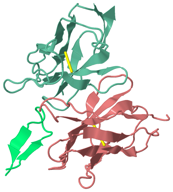
NMR StructureChain H from PDB Type:PROTEIN Length:119 aligned with HVM06_MOUSE | P01750 from UniProtKB/Swiss-Prot Length:117 Alignment length:119 117 29 39 49 59 69 79 89 99 109 | - - HVM06_MOUSE 20 HVQLQQPGAELVKPGASVKVSCKASGYTFTSYWMHWVKQRPGQGLEWIGRIHPSDSDTNYNQKFKGKATLTVDKSSSTAYMQLSSLTSEDSAVYYCAI--------------------- - SCOP domains d1qnzh_ H: Immunoglobulin heavy chain variable domain, VH SCOP domains CATH domains 1qnzH00 H:113-231 Immunoglobulins CATH domains Pfam domains -V-set-1qnzH01 H:114-210 --------------------- Pfam domains SAPs(SNPs) ----------------------------------------------------------------------------------------------------------------------- SAPs(SNPs) PROSITE ----------------------------------------------------------------------------------------------------------------------- PROSITE Transcript ----------------------------------------------------------------------------------------------------------------------- Transcript 1qnz H 113 QVQLQQSGAELVKPGASVKMSCKASGYTFTTYPIEWMKQNHGKSLEWIGNFHPYSDDTNYNEKFKGKAKLTVEKSSSTVYLEFSRLTSDDSAVYYCAIHYGSAYAMDYWGQGTSVTVSS 231 122 132 142 152 162 172 182 192 202 212 222 Chain L from PDB Type:PROTEIN Length:112 aligned with KV3AD_MOUSE | P01665 from UniProtKB/Swiss-Prot Length:111 Alignment length:112 111 10 20 30 40 50 60 70 80 90 100 110| KV3AD_MOUSE 1 DIVLTQSPASLAVSLGQRATISCKASQSVDYDGDSYMNWYQQKPGQPPKLLIYAASNLESGIPARFSGSGSGTDFTLNIHPVEEEDAATYYCQQSNEDPFTFGSGTKLEIK- - SCOP domains d1qnzl_ L: Immunoglobulin light chain kappa variable domain, VL-kappa SCOP domains CATH domains 1qnzL00 L:1-112 Immunoglobulins CATH domains Pfam domains V-set-1qnzL01 L:1-110 -- Pfam domains SAPs(SNPs) ---------------------------------------------------------------------------------------------------------------- SAPs(SNPs) PROSITE ---------------------------------------------------------------------------------------------------------------- PROSITE Transcript ---------------------------------------------------------------------------------------------------------------- Transcript 1qnz L 1 DIVLTQSPASLAVSLGQRATISCKASQSVDYDGDSYMNWYQQKPGQPPKLLIYAASNLESGIPARFSGSGSRTDFTLNIHPVEEEDAATYYCQQSNEDPFTFGSGTKLEIKR 112 10 20 30 40 50 60 70 80 90 100 110 Chain P from PDB Type:PROTEIN Length:18 aligned with Q79416_9HIV1 | Q79416 from UniProtKB/TrEMBL Length:36 Alignment length:18 18 Q79416_9HIV1 9 RKSIRIQRGPGRAFVTIG 26 SCOP domains ------------------ SCOP domains CATH domains ------------------ CATH domains Pfam domains GP120-1qnzP01 Pfam domains SAPs(SNPs) ------------------ SAPs(SNPs) PROSITE ------------------ PROSITE Transcript ------------------ Transcript 1qnz P 232 RKSIRIQRGPGRAFVTIG 249 241
|
||||||||||||||||||||
SCOP Domains (2, 2)
NMR Structure
 |
CATH Domains (1, 2)
NMR Structure
|
Pfam Domains (2, 3)
NMR Structure
|
Gene Ontology (9, 10)|
NMR Structure(hide GO term definitions) Chain H (HVM06_MOUSE | P01750)
Chain L (KV3AD_MOUSE | P01665)
Chain P (Q79416_9HIV1 | Q79416)
|
||||||||||||||||||||||||||||||||||||||||||||||||||||||||||||||||||||||||||||||||||||
Interactive Views
|
||||||||||||||||||||||||||||||||||||||||||||||||||||||||||||||||||||||||||||||||||||||||||||||||||||||||||||||||||||||||||||
Still Images
|
||||||||||||||||
Databases
|
||||||||||||||||||||||||||||||||||||||||||||||||||||||||||||||||||||||||||||||||||||||||||||||||||||||||||||||||||||||||||||||||||||||||||||||||||||||||||||||||||||||||||||||||||||||||||||||||||||||||||||||||||||
Analysis Tools
|
|||||||||||||||||||||||||||||||||||||||||||||||||||||||||||||||||||||||||||||||||||
Entries Sharing at Least One Protein Chain (UniProt ID)
Related Entries Specified in the PDB File
|
|