Show PDB file:   
         Plain Text   HTML   (compressed file size)
QuickSearch:   
by PDB,NDB,UniProt,PROSITE Code or Search Term(s)  
(-)Asym./Biol. Unit
(-)Asym./Biol. Unit - sites
collapse expand < >
Image Asym./Biol. Unit
Asym./Biol. Unit  (Jmol Viewer)
Image Asym./Biol. Unit - sites
Asym./Biol. Unit - sites  (Jmol Viewer)

(-) Description

Title :  PLASMODIUM FALCIPARUM CYCLOPHILIN COMPLEXED WITH CYCLOSPORIN A
 
Authors :  M. R. Peterson, D. R. Hall, W. N. Hunter
Date :  14 Oct 99  (Deposition) - 13 Oct 00  (Release) - 05 Jul 17  (Revision)
Method :  X-RAY DIFFRACTION
Resolution :  2.10
Chains :  Asym./Biol. Unit :  A,D
Keywords :  Isomerase-Immunosuppressant Complex, Cyclophilin-Cyclosporin Complex, Cyclosporin A, Immunosuppressant, Cyclophilin (Keyword Search: [Gene Ontology, PubMed, Web (Google))
 
Reference :  M. R. Peterson, D. R. Hall, M. Berriman, G. A. Leonard, A. H. Fairlamb, W. N. Hunter
The Three-Dimensional Structure Of A Plasmodium Falciparum Cyclophilin In Complex With The Potent Anti-Malarial Cyclosporin A
J. Mol. Biol. V. 298 123 2000
PubMed-ID: 10756109  |  Reference-DOI: 10.1006/JMBI.2000.3633
(for further references see the PDB file header)

(-) Compounds

Molecule 1 - PEPTIDYL-PROLYL CIS-TRANS ISOMERASE
    ChainsA
    EC Number5.2.1.8
    EngineeredYES
    Expression SystemESCHERICHIA COLI
    Expression System StrainBL21(DE3)
    Expression System Taxid469008
    Organism ScientificPLASMODIUM FALCIPARUM
    Organism Taxid5833
    SynonymPPIASE, ROTAMASE, CYCLOPHILIN A
 
Molecule 2 - CYCLOSPORIN A
    ChainsD
    Organism ScientificTOLYPOCLADIUM INFLATUM
    Organism Taxid29910
    SynonymCYCLOSPORINE, CICLOSPORIN, CICLOSPORINE

 Structural Features

(-) Chains, Units

  12
Asymmetric/Biological Unit AD

Summary Information (see also Sequences/Alignments below)

(-) Ligands, Modified Residues, Ions  (6, 9)

Asymmetric/Biological Unit (6, 9)
No.NameCountTypeFull Name
1ABA1Mod. Amino AcidALPHA-AMINOBUTYRIC ACID
2BMT1Mod. Amino Acid4-METHYL-4-[(E)-2-BUTENYL]-4,N-METHYL-THREONINE
3DAL1Mod. Amino AcidD-ALANINE
4MLE4Mod. Amino AcidN-METHYLLEUCINE
5MVA1Mod. Amino AcidN-METHYLVALINE
6SAR1Mod. Amino AcidSARCOSINE

(-) Sites  (1, 1)

Asymmetric Unit (1, 1)
No.NameEvidenceResiduesDescription
1AC1SOFTWAREILE A:22 , ASN A:52 , ARG A:62 , PHE A:67 , MET A:68 , GLN A:70 , GLY A:79 , ALA A:108 , ASN A:109 , ALA A:110 , GLN A:118 , PHE A:120 , TRP A:128 , LEU A:129 , HIS A:133 , GLU A:141 , HOH D:2001 , HOH D:2002 , HOH D:2003BINDING SITE FOR CHAIN D OF CYCLOSPORIN A

(-) SS Bonds  (0, 0)

(no "SS Bond" information available for 1QNG)

(-) Cis Peptide Bonds  (0, 0)

(no "Cis Peptide Bond" information available for 1QNG)

 Sequence-Structure Mapping

(-) SAPs(SNPs)/Variants  (0, 0)

(no "SAP(SNP)/Variant" information available for 1QNG)

(-) PROSITE Motifs  (0, 0)

(no "PROSITE Motif" information available for 1QNG)

(-) Exons   (0, 0)

(no "Exon" information available for 1QNG)

(-) Sequences/Alignments

Asymmetric/Biological Unit
   Reformat: Number of residues per line =  ('0' or empty: single-line sequence representation)
  Number of residues per labelling interval =   
  UniProt sequence: complete  aligned part    
   Show mapping: SCOP domains CATH domains Pfam domains Secondary structure (by author)
SAPs(SNPs) PROSITE motifs Exons
(details for a mapped element are shown in a popup box when the mouse pointer rests over it)
Chain A from PDB  Type:PROTEIN  Length:170
 aligned with Q25756_PLAFA | Q25756 from UniProtKB/TrEMBL  Length:171

    Alignment length:170
                                    11        21        31        41        51        61        71        81        91       101       111       121       131       141       151       161       171
         Q25756_PLAFA     2 SKRSKVFFDISIDNSNAGRIIFELFSDITPRTCENFRALCTGEKIGSRGKNLHYKNSIFHRIIPQFMCQGGDITNGNGSGGESIYGRSFTDENFNMKHDQPGLLSMANAGPNTNSSQFFITLVPCPWLDGKHVVFGKVIEGMNVVREMEKEGAKSGYVKRSVVITDCGEL 171
               SCOP domains d1qnga_ A: Cyclophilin (eukaryotic)                                                                                                                                        SCOP domains
               CATH domains 1qngA00 A:2-171 Cyclophilin                                                                                                                                                CATH domains
               Pfam domains ------Pro_isomerase-1qngA01 A:8-170                                                                                                                                      - Pfam domains
         Sec.struct. author ....eeeeeeee...eeeeeeeee....hhhhhhhhhhhhhh.ee.....ee........eee...eeee................................eeee...........eeee...hhhhh....eeeeeeehhhhhhhhhh...........eeeeeeeee Sec.struct. author
                 SAPs(SNPs) -------------------------------------------------------------------------------------------------------------------------------------------------------------------------- SAPs(SNPs)
                    PROSITE -------------------------------------------------------------------------------------------------------------------------------------------------------------------------- PROSITE
                 Transcript -------------------------------------------------------------------------------------------------------------------------------------------------------------------------- Transcript
                 1qng A   2 SKRSKVFFDISIDNSNAGRIIFELFSDITPRTCENFRALCTGEKIGSRGKNLHYKNSIFHRIIPQFMCQGGDITNGNGSGGESIYGRSFTDENFNMKHDQPGLLSMANAGPNTNSSQFFITLVPCPWLDGKHVVFGKVIEGMNVVREMEKEGAKSGYVKRSVVITDCGEL 171
                                    11        21        31        41        51        61        71        81        91       101       111       121       131       141       151       161       171

Chain D from PDB  Type:PROTEIN  Length:11
                                           
               SCOP domains ----------- SCOP domains
               CATH domains ----------- CATH domains
               Pfam domains ----------- Pfam domains
         Sec.struct. author ........... Sec.struct. author
                 SAPs(SNPs) ----------- SAPs(SNPs)
                    PROSITE ----------- PROSITE
                 Transcript ----------- Transcript
                 1qng D 201 xllvtaglVlA 211
                            |||||||210 
                          201-DAL||| | 
                           202-MLE|| | 
                            203-MLE| | 
                             204-MVA | 
                              205-BMT| 
                               206-ABA 
                                207-SAR
                                 208-MLE
                                   210-MLE

   Legend:   → Mismatch (orange background)
  - → Gap (green background, '-', border residues have a numbering label)
    → Modified Residue (blue background, lower-case, 'x' indicates undefined single-letter code, labelled with number + name)
  x → Chemical Group (purple background, 'x', labelled with number + name, e.g. ACE or NH2)
  extra numbering lines below/above indicate numbering irregularities and modified residue names etc., number ends below/above '|'

 Classification and Annotation

(-) SCOP Domains  (1, 1)

Asymmetric/Biological Unit

(-) CATH Domains  (1, 1)

Asymmetric/Biological Unit
(-)
Class: Mainly Beta (13760)

(-) Pfam Domains  (1, 1)

Asymmetric/Biological Unit

(-) Gene Ontology  (4, 4)

Asymmetric/Biological Unit(hide GO term definitions)
Chain A   (Q25756_PLAFA | Q25756)
molecular function
    GO:0016853    isomerase activity    Catalysis of the geometric or structural changes within one molecule. Isomerase is the systematic name for any enzyme of EC class 5.
    GO:0003755    peptidyl-prolyl cis-trans isomerase activity    Catalysis of the reaction: peptidyl-proline (omega=180) = peptidyl-proline (omega=0).
biological process
    GO:0006457    protein folding    The process of assisting in the covalent and noncovalent assembly of single chain polypeptides or multisubunit complexes into the correct tertiary structure.
    GO:0000413    protein peptidyl-prolyl isomerization    The modification of a protein by cis-trans isomerization of a proline residue.

 Visualization

(-) Interactive Views

Asymmetric/Biological Unit
  Complete Structure
    Jena3D(integrated viewing of ligand, site, SAP, PROSITE, SCOP information)
    WebMol | AstexViewer[tm]@PDBe
(Java Applets, require no local installation except for Java; loading may be slow)
    STRAP
(Java WebStart application, automatic local installation, requires Java; full application with system access!)
    RasMol
(require local installation)
    Molscript (VRML)
(requires installation of a VRML viewer; select preferred view via VRML and generate a mono or stereo PDF format file)
 
  Ligands, Modified Residues, Ions
    ABA  [ RasMol | Jena3D ]  +environment [ RasMol | Jena3D ]
    BMT  [ RasMol | Jena3D ]  +environment [ RasMol | Jena3D ]
    DAL  [ RasMol | Jena3D ]  +environment [ RasMol | Jena3D ]
    MLE  [ RasMol | Jena3D ]  +environment [ RasMol | Jena3D ]
    MVA  [ RasMol | Jena3D ]  +environment [ RasMol | Jena3D ]
    SAR  [ RasMol | Jena3D ]  +environment [ RasMol | Jena3D ]
 
  Sites
    AC1  [ RasMol ]  +environment [ RasMol ]
 
  Cis Peptide Bonds
(no "Cis Peptide Bonds" information available for 1qng)
 

(-) Still Images

Jmol
  protein: cartoon or spacefill or dots and stick; nucleic acid: cartoon and stick; ligands: spacefill; active site: stick
Molscript
  protein, nucleic acid: cartoon; ligands: spacefill; active site: ball and stick

 Databases and Analysis Tools

(-) Databases

Access by PDB/NDB ID
  1qng
    Family and Domain InformationProDom | SYSTERS
    General Structural InformationGlycoscienceDB | MMDB | NDB | OCA | PDB | PDBe | PDBj | PDBsum | PDBWiki | PQS | PROTEOPEDIA
    Orientation in MembranesOPM
    Protein SurfaceSURFACE
    Secondary StructureDSSP (structure derived) | HSSP (homology derived)
    Structural GenomicsGeneCensus
    Structural NeighboursCE | VAST
    Structure ClassificationCATH | Dali | SCOP
    Validation and Original DataBMRB Data View | BMRB Restraints Grid | EDS | PROCHECK | RECOORD | WHAT_CHECK
 
Access by UniProt ID/Accession number
  Q25756_PLAFA | Q25756
    Comparative Protein Structure ModelsModBase
    Genomic InformationEnsembl
    Protein-protein InteractionDIP
    Sequence, Family and Domain InformationInterPro | Pfam | SMART | UniProtKB/TrEMBL
 
Access by Enzyme Classificator   (EC Number)
  5.2.1.8
    General Enzyme InformationBRENDA | EC-PDB | Enzyme | IntEnz
    PathwayKEGG | MetaCyc
 
Access by Disease Identifier   (MIM ID)
  (no 'MIM ID' available)
    Disease InformationOMIM
 
Access by GenAge ID
  (no 'GenAge ID' available)
    Age Related InformationGenAge

(-) Analysis Tools

Access by PDB/NDB ID
    Domain InformationXDom
    Interatomic Contacts of Structural UnitsCSU
    Ligand-protein ContactsLPC
    Protein CavitiescastP
    Sequence and Secondary StructurePDBCartoon
    Structure AlignmentSTRAP(Java WebStart application, automatic local installation, requires Java; full application with system access!)
    Structure and Sequence BrowserSTING
 
Access by UniProt ID/Accession number
  Q25756_PLAFA | Q25756
    Protein Disorder PredictionDisEMBL | FoldIndex | GLOBPLOT (for more information see DisProt)

 Related Entries

(-) Entries Sharing at Least One Protein Chain (UniProt ID)

UniProtKB/TrEMBL
        Q25756_PLAFA | Q257561qnh

(-) Related Entries Specified in the PDB File

1bck CRYSTAL STRUCTURE OF HUMAN CYCLOPHILIN A COMPLEXED WITH CYCLOSPORIN C
1c5f CRYSTAL STRUCTURE OF THE CYCLOPHILIN-LIKE DOMAIN FROM BRUGIA MALAYI COMPLEXED WITH CYCLOSPORIN A
1csa SOLUTION STRUCTURE OF E.COLI CYCLOPHILIN (F112W) COMPLEXED WITH CYCLOSPORIN A
1cwa CRYSTAL STRUCTURE OF HUMAN CYCLOPHILIN A COMPLEXED WITH CYCLOSPORIN A
1cwb CRYSTAL STRUCTURE OF HUMAN CYCLOPHILIN A COMPLEXED WITH MODIFIED CYCLOSPORIN A AT POSITION 5
1cwc CRYSTAL STRUCTURE OF HUMAN CYCLOPHILIN A COMPLEXED WITH MODIFIED CYCLOSPORIN A AT POSITION 8
1cwf CRYSTAL STRUCTURE OF HUMAN CYCLOPHILIN A COMPLEXED WITH CYCLOSPORIN D
1cwh CRYSTAL STRUCTURE OF HUMAN CYCLOPHILIN A COMPLEXED WITH CYCLOSPORIN A MODIFIED AT POSITION 7
1cwi CRYSTAL STRUCTURE OF HUMAN CYCLOPHILIN A COMPLEXED WITH MODIFIED CYCLOSPORIN D AT POSITION 7
1cwj CRYSTAL STRUCTURE OF HUMAN CYCLOPHILIN A COMPLEXED WITH MODIFIED CYCLOSPORIN D AT POSITIONS 5 AND 7.
1cwk CRYSTAL STRUCTURE OF HUMAN CYCLOPHILIN A COMPLEXED WITH MODIFIED CYCLOSPORIN D AT POSITIONS 5 AND 7.
1cwl CRYSTAL STRUCTURE OF HUMAN CYCLOPHILIN A COMPLEXED WITH MODIFIED CYCLOSPORIN A AT POSITION 8
1cwm CRYSTAL STRUCTURE OF HUMAN CYCLOPHILIN A COMPLEXED WITH MODIFIED CYCLOSPORIN A AT POSITION 8
1cwo CRYSTAL STRUCTURE OF HUMAN CYCLOPHILIN A COMPLEXED WITH NODIFIED CYCLOSPORIN C AT POSITIONS 1, AND 9
1cya SOLUTION STRUCTURE OF HUMAN CYCLOPHILIN COMPLEXED WIYH CYCLOSPORIN A
1cyb SOLUTION STRUCTURE OF HUMAN CYCLOPHILIN COMPLEXED WITH CYCLOSPORIN A
1cyn CRYSTAL STRUCTURE OF HUMAN CYCLOPHILIN B COMPLEXED WITH MODIFIED CYCLOSPORIN A
1ikf CRYSTAL STRUCTURE OF CTCLOSPORIN-FAB COMPLEX
1m63 CRYSTAL STRUCTURE OF CALCINEURIN-CYCLOPHILIN-CYCLOSPORIN COMPLEX
1mf8 CRYSTAL STRUCTURE OF HUMAN CALCINEURIN COMPLEXED WITH HUMAN CYCLOPHILIN AND CYCLOSPORIN A
1mik CRYSTAL STRUCTURE OF HUMAN CYCLOPHILIN A COMPLEXED WITH MODIFIED CYCLOSPORIN A AT POSITION 6
1qnh CRYSTAL STRUCTURE OF PLASMODIUM FALCIPARUM CYCLOPHILIN (DOUBLE MUTANT) COMPLEXED WITH CYCLOSPORIN A
1xq7 CRYSTAL STRUCTURE OF TRYPANOSOMA CRUZI CYCLOPHILIN COMPLEXED WITH CYCLOSPORIN A
2esl CRYSTAL STRUCTURE OF HUMAN CYCLOPHILIN C COMPLEXED WITH CYCLOSPORIN A
2oju CRYSTAL STRUCTURE OF HUMAN CYCLOPHILIN J COMPLEXED WITH CYCLOSPORIN A
2poy CRYSTAL STRUCTURE OF CRYPTOSPORIDIUM PARVUM IOWA II CYCLOPHILIN A COMPLEXED WITH CYCLOSPORIN A
2rma CRYSTAL STRUCTURE OF HUMAN CYCLOPHILIN A COMPLEXED WITH CYCLOSPORIN A
2rmb CRYSTAL STRUCTURE OF HUMAN CYCLOPHILIN A COMPLEXED WITH MODIFIED CYCLOSPORIN A AT POSITION 5
2rmc CRYSTAL STRUCTURE OF MURINE CYCLOPHILIN C COMPLEXED WITH CYCLOSPORIN A
2wfj CRYSTAL STRUCTURE OF THE PPIASE DOMAIN OF HUMAN CYCLOPHILIN G COMPLEXED WITH CYCLOSPORIN A
2x2c CRYSTAL STRUCTURE OF HUMAN ACETYL-CYPA COMPLEXED WITH CYCLOSPORINE A
2x7k CRYSTAL STRUCTURE OF PPIL1 COMPLEXED WITH CYCLOSPORINE A
2z6w CRYSTAL STRUCTURE OF HUMAN CYCLOPHILIN D IN COMPLEX WITH CYCLOSPORIN A
3bo7 CRYSTAL STRUCTURE OF CYCLOSPHILIN A FROM TOXOPLASMA GONDII COMPLEXED WIT CYCLOSPORIN A
3cys SOLUTION STRUCTURE OF THE HUMAN CYCLOSPORIN A COMPLEXED WITH CYCLOSPORIN A
3eov CRYSTAL STRUCTURE OF CYCLOPHILIN FROM LEISHMANIA DONOVANI COMPLEXED WITH CYCLOSPORIN A
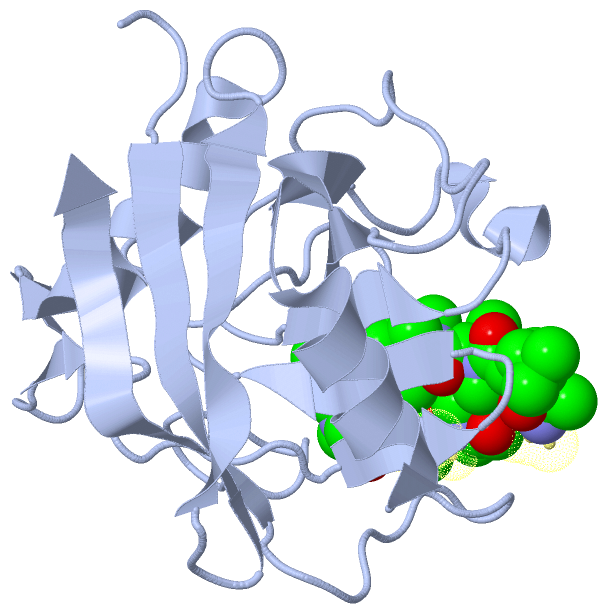
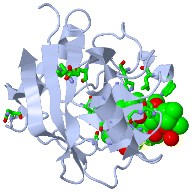