| molecular function |
|---|
| | GO:0070063 | | RNA polymerase binding | | Interacting selectively and non-covalently with an RNA polymerase molecule or complex. |
| | GO:0005518 | | collagen binding | | Interacting selectively and non-covalently with collagen, a group of fibrous proteins of very high tensile strength that form the main component of connective tissue in animals. Collagen is highly enriched in glycine (some regions are 33% glycine) and proline, occurring predominantly as 3-hydroxyproline (about 20%). |
| | GO:0016853 | | isomerase activity | | Catalysis of the geometric or structural changes within one molecule. Isomerase is the systematic name for any enzyme of EC class 5. |
| | GO:0042277 | | peptide binding | | Interacting selectively and non-covalently with peptides, any of a group of organic compounds comprising two or more amino acids linked by peptide bonds. |
| | GO:0003755 | | peptidyl-prolyl cis-trans isomerase activity | | Catalysis of the reaction: peptidyl-proline (omega=180) = peptidyl-proline (omega=0). |
| | GO:0005515 | | protein binding | | Interacting selectively and non-covalently with any protein or protein complex (a complex of two or more proteins that may include other nonprotein molecules). |
| | GO:0032403 | | protein complex binding | | Interacting selectively and non-covalently with any protein complex (a complex of two or more proteins that may include other nonprotein molecules). |
| | GO:0051082 | | unfolded protein binding | | Interacting selectively and non-covalently with an unfolded protein. |
| biological process |
|---|
| | GO:0060348 | | bone development | | The process whose specific outcome is the progression of bone over time, from its formation to the mature structure. Bone is the hard skeletal connective tissue consisting of both mineral and cellular components. |
| | GO:0061077 | | chaperone-mediated protein folding | | The process of inhibiting aggregation and assisting in the covalent and noncovalent assembly of single chain polypeptides or multisubunit complexes into the correct tertiary structure that is dependent on interaction with a chaperone. |
| | GO:0044829 | | positive regulation by host of viral genome replication | | A process in which a host organism activates or increases the frequency, rate or extent of viral genome replication. |
| | GO:0044794 | | positive regulation by host of viral process | | A process in which a host organism activates or increases the frequency, rate or extent of the release of a process being mediated by a virus with which it is infected. |
| | GO:0040018 | | positive regulation of multicellular organism growth | | Any process that activates or increases the frequency, rate or extent of growth of an organism to reach its usual body size. |
| | GO:0006457 | | protein folding | | The process of assisting in the covalent and noncovalent assembly of single chain polypeptides or multisubunit complexes into the correct tertiary structure. |
| | GO:0000413 | | protein peptidyl-prolyl isomerization | | The modification of a protein by cis-trans isomerization of a proline residue. |
| | GO:0050821 | | protein stabilization | | Any process involved in maintaining the structure and integrity of a protein and preventing it from degradation or aggregation. |
| | GO:1901873 | | regulation of post-translational protein modification | | Any process that modulates the frequency, rate or extent of post-translational protein modification. |
| cellular component |
|---|
| | GO:0005783 | | endoplasmic reticulum | | The irregular network of unit membranes, visible only by electron microscopy, that occurs in the cytoplasm of many eukaryotic cells. The membranes form a complex meshwork of tubular channels, which are often expanded into slitlike cavities called cisternae. The ER takes two forms, rough (or granular), with ribosomes adhering to the outer surface, and smooth (with no ribosomes attached). |
| | GO:0034663 | | endoplasmic reticulum chaperone complex | | A protein complex that is located in the endoplasmic reticulum and is composed of chaperone proteins, including BiP, GRP94; CaBP1, protein disulfide isomerase (PDI), ERdj3, cyclophilin B, ERp72, GRP170, UDP-glucosyltransferase, and SDF2-L1. |
| | GO:0005788 | | endoplasmic reticulum lumen | | The volume enclosed by the membranes of the endoplasmic reticulum. |
| | GO:0070062 | | extracellular exosome | | A vesicle that is released into the extracellular region by fusion of the limiting endosomal membrane of a multivesicular body with the plasma membrane. Extracellular exosomes, also simply called exosomes, have a diameter of about 40-100 nm. |
| | GO:0005925 | | focal adhesion | | Small region on the surface of a cell that anchors the cell to the extracellular matrix and that forms a point of termination of actin filaments. |
| | GO:0032991 | | macromolecular complex | | A stable assembly of two or more macromolecules, i.e. proteins, nucleic acids, carbohydrates or lipids, in which at least one component is a protein and the constituent parts function together. |
| | GO:0042470 | | melanosome | | A tissue-specific, membrane-bounded cytoplasmic organelle within which melanin pigments are synthesized and stored. Melanosomes are synthesized in melanocyte cells. |
| | GO:0016020 | | membrane | | A lipid bilayer along with all the proteins and protein complexes embedded in it an attached to it. |
| | GO:0005634 | | nucleus | | A membrane-bounded organelle of eukaryotic cells in which chromosomes are housed and replicated. In most cells, the nucleus contains all of the cell's chromosomes except the organellar chromosomes, and is the site of RNA synthesis and processing. In some species, or in specialized cell types, RNA metabolism or DNA replication may be absent. |
| | GO:0048471 | | perinuclear region of cytoplasm | | Cytoplasm situated near, or occurring around, the nucleus. |
| | GO:0005790 | | smooth endoplasmic reticulum | | The smooth endoplasmic reticulum (smooth ER or SER) has no ribosomes attached to it. The smooth ER is the recipient of the proteins synthesized in the rough ER. Those proteins to be exported are passed to the Golgi complex, the resident proteins are returned to the rough ER and the lysosomal proteins after phosphorylation of their mannose residues are passed to the lysosomes. Glycosylation of the glycoproteins also continues. The smooth ER is the site of synthesis of lipids, including the phospholipids. The membranes of the smooth ER also contain enzymes that catalyze a series of reactions to detoxify both lipid-soluble drugs and harmful products of metabolism. Large quantities of certain compounds such as phenobarbital cause an increase in the amount of the smooth ER. |
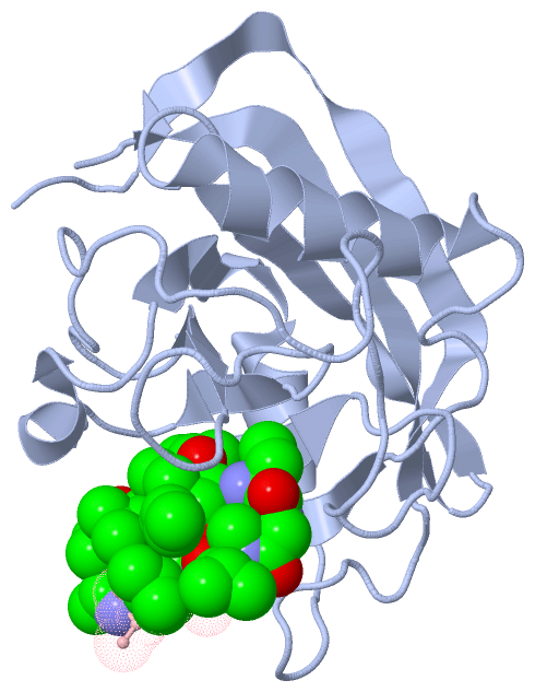
 Description
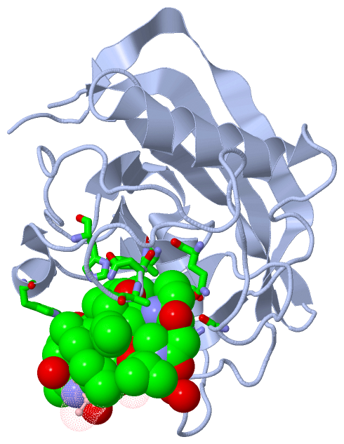