|
|
|
|
 Description
|
|
Compounds
|
||||||||||||||||||||||||||||||||||||||||||||||||
Chains, Units
Summary Information (see also Sequences/Alignments below) |
Ligands, Modified Residues, Ions (0, 0)| (no "Ligand,Modified Residues,Ions" information available for 1P4W) |
Sites (0, 0)| (no "Site" information available for 1P4W) |
SS Bonds (0, 0)| (no "SS Bond" information available for 1P4W) |
Cis Peptide Bonds (0, 0)| (no "Cis Peptide Bond" information available for 1P4W) |
SAPs(SNPs)/Variants (0, 0)| (no "SAP(SNP)/Variant" information available for 1P4W) |
PROSITE Motifs (0, 0)| (no "PROSITE Motif" information available for 1P4W) |
Exons (0, 0)| (no "Exon" information available for 1P4W) |
Sequences/Alignments
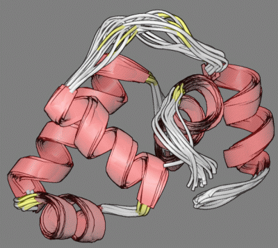
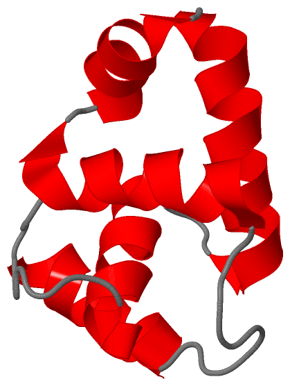
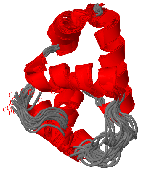
NMR StructureChain A from PDB Type:PROTEIN Length:87 aligned with P96320_ERWAM | P96320 from UniProtKB/TrEMBL Length:215 Alignment length:87 138 148 158 168 178 188 198 208 P96320_ERWAM 129 YTPESVAKLLEKISAGGYGDKRLSPKESEVLRLFAEGFLVTEIAKKLNRSIKTISSQKKSAMMKLGVDNDIALLNYLSSVSMTPVDK 215 SCOP domains d1p4wa_ A: Transcriptional regulator RcsB SCOP domains CATH domains 1p4wA00 A:129-215 'winged helix' repressor DNA binding domain CATH domains Pfam domains -------------------GerE-1p4wA01 A:148-205 ---------- Pfam domains SAPs(SNPs) --------------------------------------------------------------------------------------- SAPs(SNPs) PROSITE --------------------------------------------------------------------------------------- PROSITE Transcript --------------------------------------------------------------------------------------- Transcript 1p4w A 129 YTPESVAKLLEKISAGGYGDKRLSPKESEVLRLFAEGFLVTEIAKKLNRSIKTISSQKKSAMMKLGVDNDIALLNYLSSVSMTPVDK 215 138 148 158 168 178 188 198 208
|
||||||||||||||||||||
SCOP Domains (1, 1)
NMR Structure
 |
CATH Domains (1, 1)
NMR Structure
|
Pfam Domains (1, 1)
NMR Structure
|
Gene Ontology (6, 6)|
NMR Structure(hide GO term definitions) Chain A (P96320_ERWAM | P96320)
|
||||||||||||||||||||||||||||||||||||||||||||||||||||||
Interactive Views
|
||||||||||||||||||||||||||||||||||||||||||||||||||||||||||||||||||||||||||||||||||||||||||||||||||||||||||||||||||||
Still Images
|
||||||||||||||||
Databases
|
||||||||||||||||||||||||||||||||||||||||||||||||||||||||||||||||||||||||||||||||||||||||||||||||||||||||||||||||||||||||||||||||||||||||||||||||||||||||||||||||
Analysis Tools
|
|||||||||||||||||||||||||||||||||||||||||||||||||||||||||||||
Entries Sharing at Least One Protein Chain (UniProt ID)
Related Entries Specified in the PDB File
|
|