|
|
|
|
 Description
|
|
Compounds
|
||||||||||||||||||||||||||||||||||||||||||||
Chains, Units
Summary Information (see also Sequences/Alignments below) |
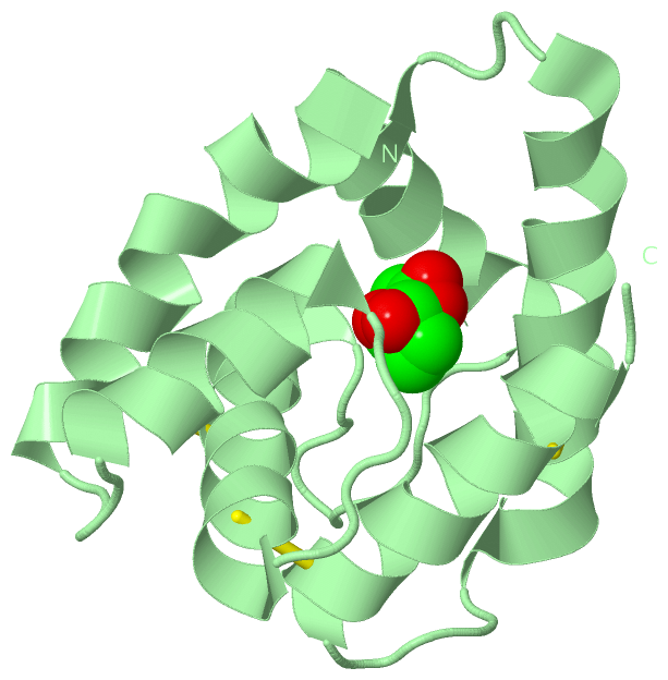
Ligands, Modified Residues, Ions (2, 4)
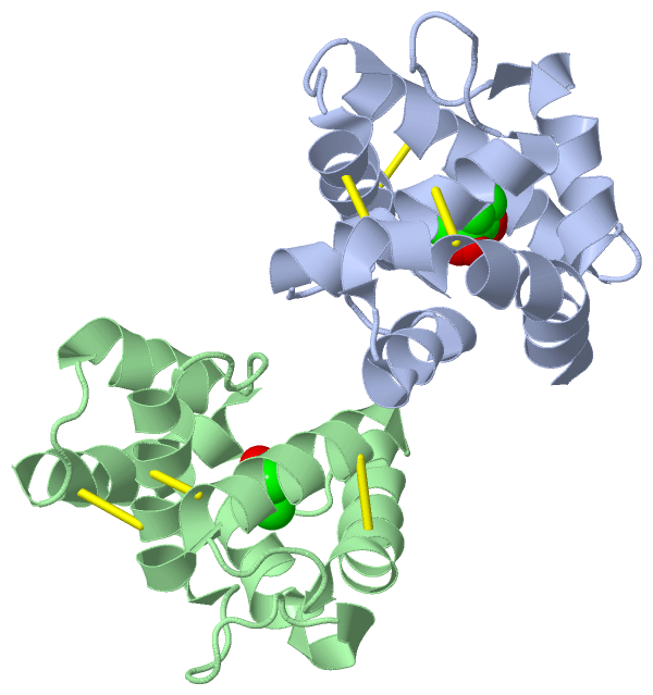
Asymmetric Unit (2, 4)
|
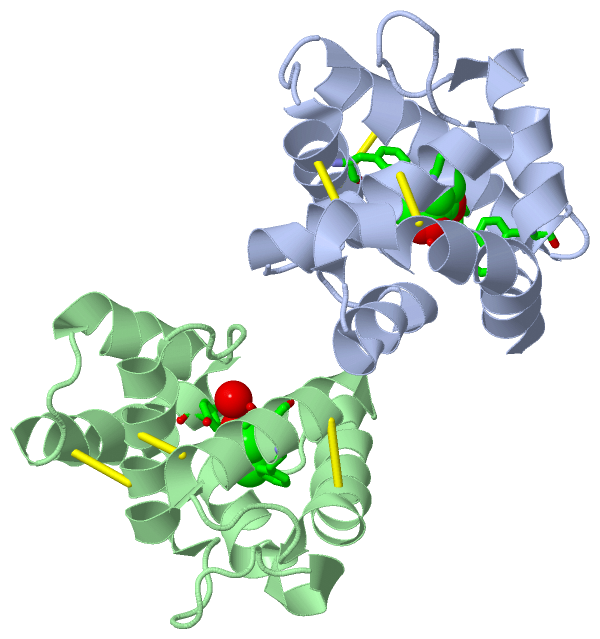
Sites (4, 4)
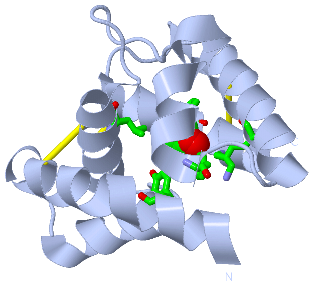
Asymmetric Unit (4, 4)
|
SS Bonds (6, 6)
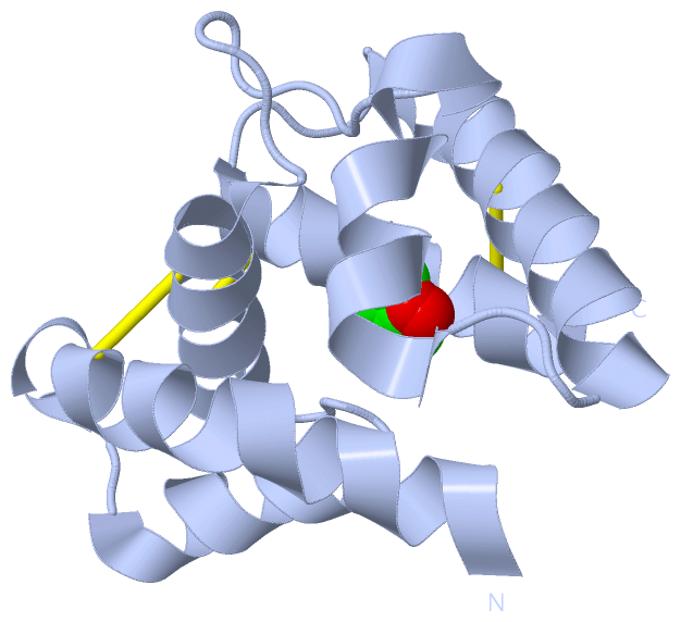
Asymmetric Unit
|
||||||||||||||||||||||||||||
Cis Peptide Bonds (0, 0)| (no "Cis Peptide Bond" information available for 1P28) |
SAPs(SNPs)/Variants (0, 0)| (no "SAP(SNP)/Variant" information available for 1P28) |
PROSITE Motifs (0, 0)| (no "PROSITE Motif" information available for 1P28) |
Exons (0, 0)| (no "Exon" information available for 1P28) |
Sequences/Alignments
Asymmetric UnitChain A from PDB Type:PROTEIN Length:119 aligned with Q8MTC1_RHYMA | Q8MTC1 from UniProtKB/TrEMBL Length:137 Alignment length:119 27 37 47 57 67 77 87 97 107 117 127 Q8MTC1_RHYMA 18 ADSTQSYKDAMGPLVRECMGSVSATEDDFKTVLNRNPLESRTAQCLLACALDKVGLISPEGAIYTGDDLMPVMNRLYGFNDFKTVMKAKAVNDCANQVNGAYPDRCDLIKNFTDCVRNS 136 SCOP domains d1p28a_ A: Pheromone binding protein PBPLma SCOP domains CATH domains 1p28A00 A:-1-117 [code=1.10.238.20, no name defined] CATH domains Pfam domains ----------------------------------------------------------------------------------------------------------------------- Pfam domains SAPs(SNPs) ----------------------------------------------------------------------------------------------------------------------- SAPs(SNPs) PROSITE ----------------------------------------------------------------------------------------------------------------------- PROSITE Transcript ----------------------------------------------------------------------------------------------------------------------- Transcript 1p28 A -1 NSSTQSYKDAMGPLVRECMGSVSATEDDFKTVLNRNPLESRTAQCLLACALDKVGLISPEGAIYTGDDLMPVMNRLYGFNDFKTVMKAKAVNDCANQVNGAYPDRCDLIKNFTDCVRNS 117 8 18 28 38 48 58 68 78 88 98 108 Chain B from PDB Type:PROTEIN Length:117 aligned with Q8MTC1_RHYMA | Q8MTC1 from UniProtKB/TrEMBL Length:137 Alignment length:117 30 40 50 60 70 80 90 100 110 120 130 Q8MTC1_RHYMA 21 TQSYKDAMGPLVRECMGSVSATEDDFKTVLNRNPLESRTAQCLLACALDKVGLISPEGAIYTGDDLMPVMNRLYGFNDFKTVMKAKAVNDCANQVNGAYPDRCDLIKNFTDCVRNSY 137 SCOP domains d1p28b_ B: Pheromone binding protein PBPLma SCOP domains CATH domains 1p28B00 B:2-118 [code=1.10.238.20, no name defined] CATH domains Pfam domains (1) PBP_GOBP-1p28B01 B:2-118 Pfam domains (1) Pfam domains (2) PBP_GOBP-1p28B02 B:2-118 Pfam domains (2) SAPs(SNPs) --------------------------------------------------------------------------------------------------------------------- SAPs(SNPs) PROSITE --------------------------------------------------------------------------------------------------------------------- PROSITE Transcript --------------------------------------------------------------------------------------------------------------------- Transcript 1p28 B 2 TQSYKDAMGPLVRECMGSVSATEDDFKTVLNRNPLESRTAQCLLACALDKVGLISPEGAIYTGDDLMPVMNRLYGFNDFKTVMKAKAVNDCANQVNGAYPDRCDLIKNFTDCVRNSY 118 11 21 31 41 51 61 71 81 91 101 111
|
||||||||||||||||||||
SCOP Domains (1, 2)
Asymmetric Unit
 |
CATH Domains (1, 2)
Asymmetric Unit
|
Pfam Domains (1, 2)
Asymmetric Unit
|
Gene Ontology (1, 1)|
Asymmetric Unit(hide GO term definitions) Chain A,B (Q8MTC1_RHYMA | Q8MTC1)
|
||||||||||||
Interactive Views
|
|||||||||||||||||||||||||||||||||||||||||||||||||||||||||||||||||||||||||||||||||||||||||||||||||||||||||||||||||||||||||||||||||||||||||||||||||||||||||||||||||||||||||
Still Images
|
||||||||||||||||
Databases
|
||||||||||||||||||||||||||||||||||||||||||||||||||||||||||||||||||||||||||||||||||||||||||||||||||||||||||||||||||||||||||||||||||||||||||||||||||||||||||||||||
Analysis Tools
|
|||||||||||||||||||||||||||||||||||||||||||||||||||||||||||||
Entries Sharing at Least One Protein Chain (UniProt ID)
Related Entries Specified in the PDB File
|
|