Show PDB file:   
         Plain Text   HTML   (compressed file size)
QuickSearch:   
by PDB,NDB,UniProt,PROSITE Code or Search Term(s)  
(-)Asym.Unit - manually
(-)Asymmetric Unit
(-)Asym. Unit - sites
(-)Biological Unit 1
(-)Biol. Unit 1 - sites
(-)Biological Unit 2
collapse expand < >
Image Asym.Unit - manually
Asym.Unit - manually  (Jmol Viewer)
Image Asymmetric Unit
Asymmetric Unit  (Jmol Viewer)
Image Asym. Unit - sites
Asym. Unit - sites  (Jmol Viewer)
Image Biological Unit 1
Biological Unit 1  (Jmol Viewer)
Image Biol. Unit 1 - sites
Biol. Unit 1 - sites  (Jmol Viewer)
Image Biological Unit 2
Biological Unit 2  (Jmol Viewer)

(-) Description

Title :  THE CRYSTAL STRUCTURE OF A PHEROMONE BINDING PROTEIN FROM THE COCKROACH LEUCOPHAEA MADERAE REVEALS A NEW MECHANISM OF PHEROMONE BINDING
 
Authors :  A. Lartigue, A. Gruez, S. Spinelli, S. Riviere, R. Brossut, M. Tegoni, C. Cambillau
Date :  13 Mar 03  (Deposition) - 05 Aug 03  (Release) - 13 Jul 11  (Revision)
Method :  X-RAY DIFFRACTION
Resolution :  1.70
Chains :  Asym. Unit :  A,B
Biol. Unit 1:  A  (1x)
Biol. Unit 2:  B  (1x)
Keywords :  Pheromone Binding Protein, Apo-Form, 6 Alpha Helix, Transport Protein (Keyword Search: [Gene Ontology, PubMed, Web (Google))
 
Reference :  A. Lartigue, A. Gruez, S. Spinelli, S. Riviere, R. Brossut, M. Tegoni, C. Cambillau
The Crystal Structure Of A Cockroach Pheromone-Binding Protein Suggests A New Ligand Binding And Release Mechanism
J. Biol. Chem. V. 278 30213 2003
PubMed-ID: 12766173  |  Reference-DOI: 10.1074/JBC.M304688200

(-) Compounds

Molecule 1 - PHEROMONE BINDING PROTEIN
    ChainsA, B
    EngineeredYES
    Expression SystemESCHERICHIA COLI BL21(DE3)
    Expression System PlasmidPET22B+
    Expression System StrainBL21(DE3)
    Expression System Taxid469008
    Expression System Vector TypePLASMID
    Organism CommonMADEIRA COCKROACH
    Organism ScientificLEUCOPHAEA MADERAE
    Organism Taxid6988

 Structural Features

(-) Chains, Units

  12
Asymmetric Unit AB
Biological Unit 1 (1x)A 
Biological Unit 2 (1x) B

Summary Information (see also Sequences/Alignments below)

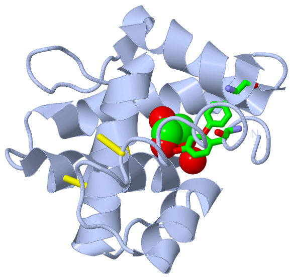
(-) Ligands, Modified Residues, Ions  (1, 2)

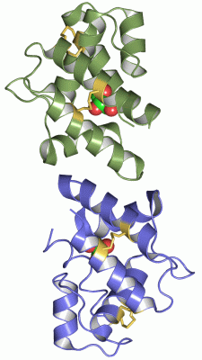
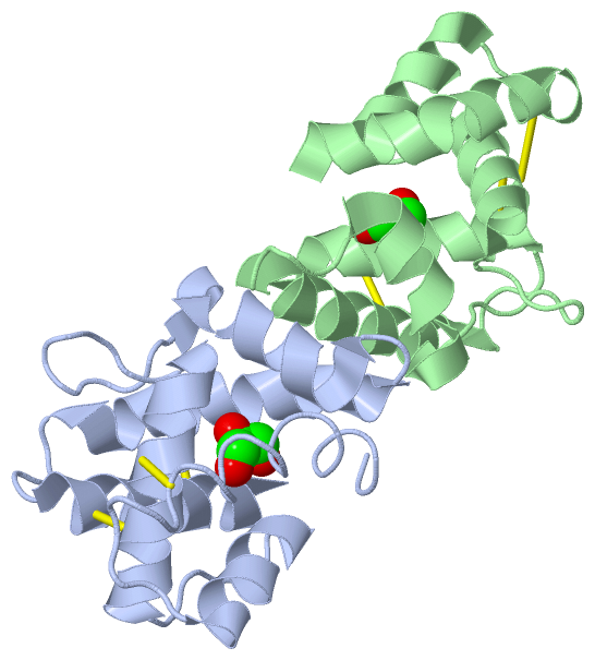
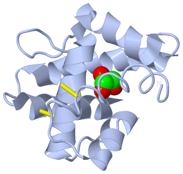
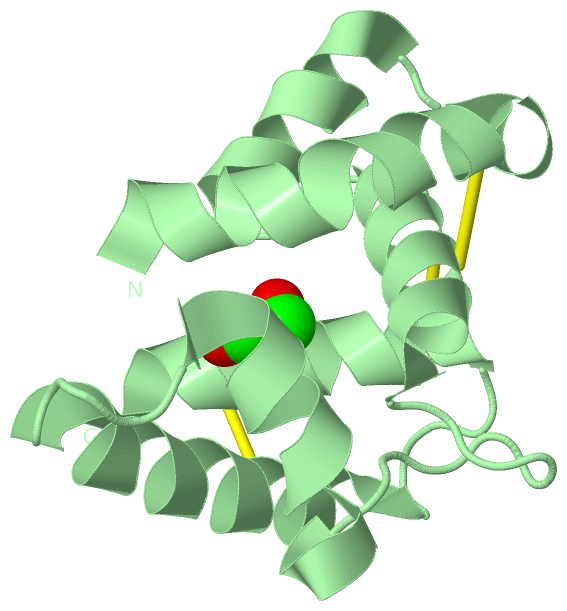
Asymmetric Unit (1, 2)
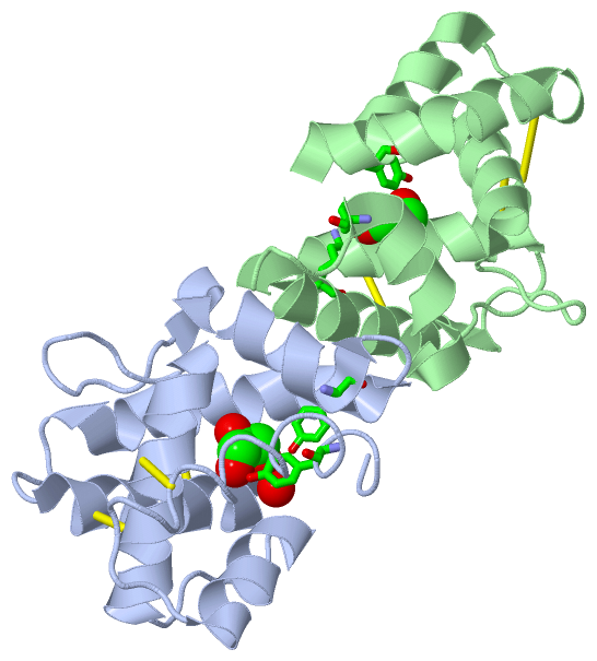
No.NameCountTypeFull Name
1GOL2Ligand/IonGLYCEROL
Biological Unit 1 (1, 1)
No.NameCountTypeFull Name
1GOL1Ligand/IonGLYCEROL
Biological Unit 2 (1, 1)
No.NameCountTypeFull Name
1GOL1Ligand/IonGLYCEROL

(-) Sites  (2, 2)

Asymmetric Unit (2, 2)
No.NameEvidenceResiduesDescription
1AC1SOFTWARETYR B:5 , TYR B:75 , LYS B:85BINDING SITE FOR RESIDUE GOL B 250
2AC2SOFTWARETYR A:5 , TYR A:75 , HOH A:349BINDING SITE FOR RESIDUE GOL A 251

(-) SS Bonds  (6, 6)

Asymmetric Unit
No.Residues
1A:16 -A:47
2A:43 -A:104
3A:92 -A:113
4B:16 -B:47
5B:43 -B:104
6B:92 -B:113

(-) Cis Peptide Bonds  (0, 0)

(no "Cis Peptide Bond" information available for 1ORG)

 Sequence-Structure Mapping

(-) SAPs(SNPs)/Variants  (0, 0)

(no "SAP(SNP)/Variant" information available for 1ORG)

(-) PROSITE Motifs  (0, 0)

(no "PROSITE Motif" information available for 1ORG)

(-) Exons   (0, 0)

(no "Exon" information available for 1ORG)

(-) Sequences/Alignments

Asymmetric Unit
   Reformat: Number of residues per line =  ('0' or empty: single-line sequence representation)
  Number of residues per labelling interval =   
  UniProt sequence: complete  aligned part    
   Show mapping: SCOP domains CATH domains Pfam domains Secondary structure (by author)
SAPs(SNPs) PROSITE motifs Exons
(details for a mapped element are shown in a popup box when the mouse pointer rests over it)
Chain A from PDB  Type:PROTEIN  Length:118
 aligned with Q8MTC1_RHYMA | Q8MTC1 from UniProtKB/TrEMBL  Length:137

    Alignment length:118
                                    29        39        49        59        69        79        89        99       109       119       129        
         Q8MTC1_RHYMA    20 STQSYKDAMGPLVRECMGSVSATEDDFKTVLNRNPLESRTAQCLLACALDKVGLISPEGAIYTGDDLMPVMNRLYGFNDFKTVMKAKAVNDCANQVNGAYPDRCDLIKNFTDCVRNSY 137
               SCOP domains d1orga_ A: Pheromone binding protein PBPLma                                                                            SCOP domains
               CATH domains 1orgA00 A:1-118  [code=1.10.238.20, no name defined]                                                                   CATH domains
               Pfam domains ---------------------------------------------------------------------------------------------------------------------- Pfam domains
         Sec.struct. author ......................hhhhhhhhhh.....hhhhhhhhhhhhhhhh.........hhhhhhhhhhhhhh..hhhhhhhhhhhhhhhhhhh....hhhhhhhhhhhhhhhh. Sec.struct. author
                 SAPs(SNPs) ---------------------------------------------------------------------------------------------------------------------- SAPs(SNPs)
                    PROSITE ---------------------------------------------------------------------------------------------------------------------- PROSITE
                 Transcript ---------------------------------------------------------------------------------------------------------------------- Transcript
                 1org A   1 STQSYKDAMGPLVRECMGSVSATEDDFKTVLNRNPLESRTAQCLLACALDKVGLISPEGAIYTGDDLMPVMNRLYGFNDFKTVMKAKAVNDCANQVNGAYPDRCDLIKNFTDCVRNSY 118
                                    10        20        30        40        50        60        70        80        90       100       110        

Chain B from PDB  Type:PROTEIN  Length:118
 aligned with Q8MTC1_RHYMA | Q8MTC1 from UniProtKB/TrEMBL  Length:137

    Alignment length:118
                                    29        39        49        59        69        79        89        99       109       119       129        
         Q8MTC1_RHYMA    20 STQSYKDAMGPLVRECMGSVSATEDDFKTVLNRNPLESRTAQCLLACALDKVGLISPEGAIYTGDDLMPVMNRLYGFNDFKTVMKAKAVNDCANQVNGAYPDRCDLIKNFTDCVRNSY 137
               SCOP domains d1orgb_ B: Pheromone binding protein PBPLma                                                                            SCOP domains
               CATH domains 1orgB00 B:1-118  [code=1.10.238.20, no name defined]                                                                   CATH domains
           Pfam domains (1) PBP_GOBP-1orgB01 B:1-118                                                                                               Pfam domains (1)
           Pfam domains (2) PBP_GOBP-1orgB02 B:1-118                                                                                               Pfam domains (2)
         Sec.struct. author hhhhhhhhhhhhhhhhhhhhh.hhhhhhhhhh.....hhhhhhhhhhhhhhhh.........hhhhhhhhhhhhh....hhhhhhhhhhhhhhhhhh....hhhhhhhhhhhhhhhh. Sec.struct. author
                 SAPs(SNPs) ---------------------------------------------------------------------------------------------------------------------- SAPs(SNPs)
                    PROSITE ---------------------------------------------------------------------------------------------------------------------- PROSITE
                 Transcript ---------------------------------------------------------------------------------------------------------------------- Transcript
                 1org B   1 STQSYKDAMGPLVRECMGSVSATEDDFKTVLNRNPLESRTAQCLLACALDKVGLISPEGAIYTGDDLMPVMNRLYGFNDFKTVMKAKAVNDCANQVNGAYPDRCDLIKNFTDCVRNSY 118
                                    10        20        30        40        50        60        70        80        90       100       110        

   Legend:   → Mismatch (orange background)
  - → Gap (green background, '-', border residues have a numbering label)
    → Modified Residue (blue background, lower-case, 'x' indicates undefined single-letter code, labelled with number + name)
  x → Chemical Group (purple background, 'x', labelled with number + name, e.g. ACE or NH2)
  extra numbering lines below/above indicate numbering irregularities and modified residue names etc., number ends below/above '|'

 Classification and Annotation

(-) SCOP Domains  (1, 2)

Asymmetric Unit

(-) CATH Domains  (1, 2)

Asymmetric Unit

(-) Pfam Domains  (1, 2)

Asymmetric Unit

(-) Gene Ontology  (1, 1)

Asymmetric Unit(hide GO term definitions)
Chain A,B   (Q8MTC1_RHYMA | Q8MTC1)
molecular function
    GO:0005549    odorant binding    Interacting selectively and non-covalently with an odorant, any substance capable of stimulating the sense of smell.

 Visualization

(-) Interactive Views

Asymmetric Unit
  Complete Structure
    Jena3D(integrated viewing of ligand, site, SAP, PROSITE, SCOP information)
    WebMol | AstexViewer[tm]@PDBe
(Java Applets, require no local installation except for Java; loading may be slow)
    STRAP
(Java WebStart application, automatic local installation, requires Java; full application with system access!)
    RasMol
(require local installation)
    Molscript (VRML)
(requires installation of a VRML viewer; select preferred view via VRML and generate a mono or stereo PDF format file)
 
  Ligands, Modified Residues, Ions
    GOL  [ RasMol | Jena3D ]  +environment [ RasMol | Jena3D ]
 
  Sites
    AC1  [ RasMol ]  +environment [ RasMol ]
    AC2  [ RasMol ]  +environment [ RasMol ]
 
  Cis Peptide Bonds
(no "Cis Peptide Bonds" information available for 1org)
 
Biological Units
  Complete Structure
    Biological Unit 1  [ Jena3D ]
    Biological Unit 2  [ Jena3D ]

(-) Still Images

Jmol
  protein: cartoon or spacefill or dots and stick; nucleic acid: cartoon and stick; ligands: spacefill; active site: stick
Molscript
  protein, nucleic acid: cartoon; ligands: spacefill; active site: ball and stick

 Databases and Analysis Tools

(-) Databases

Access by PDB/NDB ID
  1org
    Family and Domain InformationProDom | SYSTERS
    General Structural InformationGlycoscienceDB | MMDB | NDB | OCA | PDB | PDBe | PDBj | PDBsum | PDBWiki | PQS | PROTEOPEDIA
    Orientation in MembranesOPM
    Protein SurfaceSURFACE
    Secondary StructureDSSP (structure derived) | HSSP (homology derived)
    Structural GenomicsGeneCensus
    Structural NeighboursCE | VAST
    Structure ClassificationCATH | Dali | SCOP
    Validation and Original DataBMRB Data View | BMRB Restraints Grid | EDS | PROCHECK | RECOORD | WHAT_CHECK
 
Access by UniProt ID/Accession number
  Q8MTC1_RHYMA | Q8MTC1
    Comparative Protein Structure ModelsModBase
    Genomic InformationEnsembl
    Protein-protein InteractionDIP
    Sequence, Family and Domain InformationInterPro | Pfam | SMART | UniProtKB/TrEMBL
 
Access by Enzyme Classificator   (EC Number)
  (no 'Enzyme Classificator' available)
    General Enzyme InformationBRENDA | EC-PDB | Enzyme | IntEnz
    PathwayKEGG | MetaCyc
 
Access by Disease Identifier   (MIM ID)
  (no 'MIM ID' available)
    Disease InformationOMIM
 
Access by GenAge ID
  (no 'GenAge ID' available)
    Age Related InformationGenAge

(-) Analysis Tools

Access by PDB/NDB ID
    Domain InformationXDom
    Interatomic Contacts of Structural UnitsCSU
    Ligand-protein ContactsLPC
    Protein CavitiescastP
    Sequence and Secondary StructurePDBCartoon
    Structure AlignmentSTRAP(Java WebStart application, automatic local installation, requires Java; full application with system access!)
    Structure and Sequence BrowserSTING
 
Access by UniProt ID/Accession number
  Q8MTC1_RHYMA | Q8MTC1
    Protein Disorder PredictionDisEMBL | FoldIndex | GLOBPLOT (for more information see DisProt)

 Related Entries

(-) Entries Sharing at Least One Protein Chain (UniProt ID)

UniProtKB/TrEMBL
        Q8MTC1_RHYMA | Q8MTC11ow4 1p28

(-) Related Entries Specified in the PDB File

1ow4 CRYSTAL STRUCTURE OF A PHEROMONE BINDING PROTEIN FROM THE COCKROACH LEUCOPHAEA MADERAE IN COMPLEX WITH THE FLUORESCENT REPORTER ANS (1-ANILINONAPHTALENE-8-SULFONIC ACID)
1p28 THE CRYSTAL STRUCTURE OF A PHEROMONE BINDING PROTEIN FROM THE COCKROACH LEUCOPHAEA MADERAE IN COMPLEX WITH A COMPONENT OF THE PHEROMONAL BLEND: 3-HYDROXY-BUTAN-2-ONE