|
|
|
|
 Description
|
|
Compounds
|
||||||||||||||||||||||||||||||||||||
Chains, Units
Summary Information (see also Sequences/Alignments below) |
Ligands, Modified Residues, Ions (0, 0)| (no "Ligand,Modified Residues,Ions" information available for 1OFT) |
Sites (0, 0)| (no "Site" information available for 1OFT) |
SS Bonds (0, 0)| (no "SS Bond" information available for 1OFT) |
Cis Peptide Bonds (4, 4)
Asymmetric/Biological Unit
|
||||||||||||||||||||
SAPs(SNPs)/Variants (0, 0)| (no "SAP(SNP)/Variant" information available for 1OFT) |
PROSITE Motifs (0, 0)| (no "PROSITE Motif" information available for 1OFT) |
Exons (0, 0)| (no "Exon" information available for 1OFT) |
Sequences/Alignments
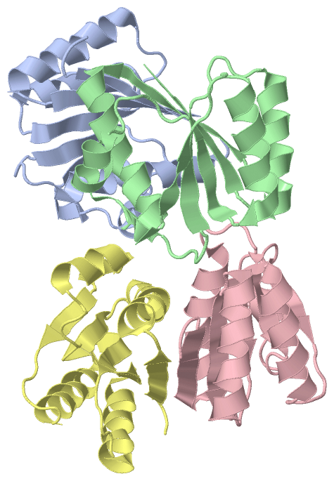
Asymmetric/Biological UnitChain A from PDB Type:PROTEIN Length:119 aligned with SULA_PSEAE | Q9HZJ8 from UniProtKB/Swiss-Prot Length:161 Alignment length:119 52 62 72 82 92 102 112 122 132 142 152 SULA_PSEAE 43 PAAFSELSLSGLPGHCLTLLAPILRELSEEQDARWLTLIAPPASLTHEWLRRAGLNRERILLLQAKDNAAALALSCEALRLGRSHTVVSWLEPLSRAARKQLSRAAQLGQAQSLNIRLG 161 SCOP domains d1ofta_ A: Hypothetical protein PA3008 SCOP domains CATH domains 1oftA00 A:43-161 P-loop containing nucleotide triphosphate hydrolases CATH domains Pfam domains ----------------------------------------------------------------------------------------------------------------------- Pfam domains SAPs(SNPs) ----------------------------------------------------------------------------------------------------------------------- SAPs(SNPs) PROSITE ----------------------------------------------------------------------------------------------------------------------- PROSITE Transcript ----------------------------------------------------------------------------------------------------------------------- Transcript 1oft A 43 PAAFSELSLSGLPGHCLTLLAPILRELSEEQDARWLTLIAPPASLTHEWLRRAGLNRERILLLQAKDNAAALALSCEALRLGRSHTVVSWLEPLSRAARKQLSRAAQLGQAQSLNIRLG 161 52 62 72 82 92 102 112 122 132 142 152 Chain B from PDB Type:PROTEIN Length:119 aligned with SULA_PSEAE | Q9HZJ8 from UniProtKB/Swiss-Prot Length:161 Alignment length:119 52 62 72 82 92 102 112 122 132 142 152 SULA_PSEAE 43 PAAFSELSLSGLPGHCLTLLAPILRELSEEQDARWLTLIAPPASLTHEWLRRAGLNRERILLLQAKDNAAALALSCEALRLGRSHTVVSWLEPLSRAARKQLSRAAQLGQAQSLNIRLG 161 SCOP domains d1oftb_ B: Hypothetical protein PA3008 SCOP domains CATH domains 1oftB00 B:43-161 P-loop containing nucleotide triphosphate hydrolases CATH domains Pfam domains ----------------------------------------------------------------------------------------------------------------------- Pfam domains SAPs(SNPs) ----------------------------------------------------------------------------------------------------------------------- SAPs(SNPs) PROSITE ----------------------------------------------------------------------------------------------------------------------- PROSITE Transcript ----------------------------------------------------------------------------------------------------------------------- Transcript 1oft B 43 PAAFSELSLSGLPGHCLTLLAPILRELSEEQDARWLTLIAPPASLTHEWLRRAGLNRERILLLQAKDNAAALALSCEALRLGRSHTVVSWLEPLSRAARKQLSRAAQLGQAQSLNIRLG 161 52 62 72 82 92 102 112 122 132 142 152 Chain C from PDB Type:PROTEIN Length:119 aligned with SULA_PSEAE | Q9HZJ8 from UniProtKB/Swiss-Prot Length:161 Alignment length:119 52 62 72 82 92 102 112 122 132 142 152 SULA_PSEAE 43 PAAFSELSLSGLPGHCLTLLAPILRELSEEQDARWLTLIAPPASLTHEWLRRAGLNRERILLLQAKDNAAALALSCEALRLGRSHTVVSWLEPLSRAARKQLSRAAQLGQAQSLNIRLG 161 SCOP domains d1oftc_ C: Hypothetical protein PA3008 SCOP domains CATH domains 1oftC00 C:43-161 P-loop containing nucleotide triphosphate hydrolases CATH domains Pfam domains ----------------------------------------------------------------------------------------------------------------------- Pfam domains SAPs(SNPs) ----------------------------------------------------------------------------------------------------------------------- SAPs(SNPs) PROSITE ----------------------------------------------------------------------------------------------------------------------- PROSITE Transcript ----------------------------------------------------------------------------------------------------------------------- Transcript 1oft C 43 PAAFSELSLSGLPGHCLTLLAPILRELSEEQDARWLTLIAPPASLTHEWLRRAGLNRERILLLQAKDNAAALALSCEALRLGRSHTVVSWLEPLSRAARKQLSRAAQLGQAQSLNIRLG 161 52 62 72 82 92 102 112 122 132 142 152 Chain D from PDB Type:PROTEIN Length:119 aligned with SULA_PSEAE | Q9HZJ8 from UniProtKB/Swiss-Prot Length:161 Alignment length:119 52 62 72 82 92 102 112 122 132 142 152 SULA_PSEAE 43 PAAFSELSLSGLPGHCLTLLAPILRELSEEQDARWLTLIAPPASLTHEWLRRAGLNRERILLLQAKDNAAALALSCEALRLGRSHTVVSWLEPLSRAARKQLSRAAQLGQAQSLNIRLG 161 SCOP domains d1oftd_ D: Hypothetical protein PA3008 SCOP domains CATH domains 1oftD00 D:43-161 P-loop containing nucleotide triphosphate hydrolases CATH domains Pfam domains (1) SulA-1oftD01 D:43-134 --------------------------- Pfam domains (1) Pfam domains (2) SulA-1oftD02 D:43-134 --------------------------- Pfam domains (2) Pfam domains (3) SulA-1oftD03 D:43-134 --------------------------- Pfam domains (3) Pfam domains (4) SulA-1oftD04 D:43-134 --------------------------- Pfam domains (4) SAPs(SNPs) ----------------------------------------------------------------------------------------------------------------------- SAPs(SNPs) PROSITE ----------------------------------------------------------------------------------------------------------------------- PROSITE Transcript ----------------------------------------------------------------------------------------------------------------------- Transcript 1oft D 43 PAAFSELSLSGLPGHCLTLLAPILRELSEEQDARWLTLIAPPASLTHEWLRRAGLNRERILLLQAKDNAAALALSCEALRLGRSHTVVSWLEPLSRAARKQLSRAAQLGQAQSLNIRLG 161 52 62 72 82 92 102 112 122 132 142 152
|
||||||||||||||||||||
SCOP Domains (1, 4)
Asymmetric/Biological Unit
 |
CATH Domains (1, 4)
Asymmetric/Biological Unit
|
Pfam Domains (1, 4)
Asymmetric/Biological Unit
|
Gene Ontology (7, 7)|
Asymmetric/Biological Unit(hide GO term definitions) Chain A,B,C,D (SULA_PSEAE | Q9HZJ8)
|
||||||||||||||||||||||||||||||||||||||||||||||||||||||
Interactive Views
|
||||||||||||||||||||||||||||||||||||||||||||||||||||||||||||||||||||||||||||||||||||||||||||||||||||||||||||||||||||||||||||||||||||||||||
Still Images
|
||||||||||||||||
Databases
|
||||||||||||||||||||||||||||||||||||||||||||||||||||||||||||||||||||||||||||||||||||||||||||||||||||||||||||||||||||||||||||||||||||||||||||||||||||||||||||||||
Analysis Tools
|
|||||||||||||||||||||||||||||||||||||||||||||||||||||||||||||
Entries Sharing at Least One Protein Chain (UniProt ID)
Related Entries Specified in the PDB File
|
|