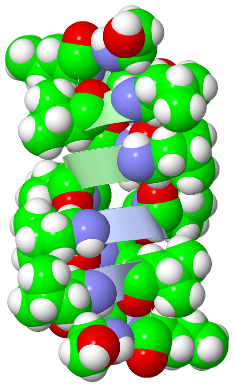
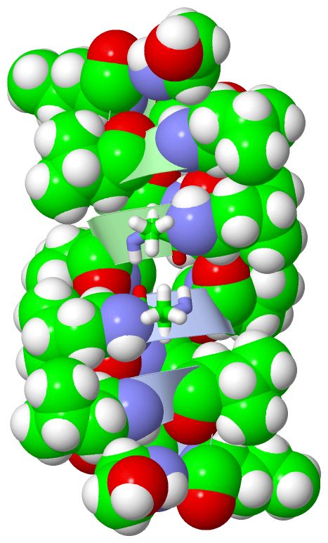
Show PDB file:   
         Plain Text   HTML   (compressed file size)
QuickSearch:   
by PDB,NDB,UniProt,PROSITE Code or Search Term(s)  
(-)NMR Structure - model 1
(-)NMR Structure - model 1, sites
(-)NMR Structure - all models
collapse expand < >
Image NMR Structure - model 1
NMR Structure - model 1  (Jmol Viewer)
Image NMR Structure - model 1, sites
NMR Structure - model 1, sites  (Jmol Viewer)
Image NMR Structure - all models
NMR Structure - all models  (Jmol Viewer)

(-) Description

Title :  G15-GRAMICIDIN A IN SODIUM DODECYL SULFATE MICELLES (NMR)
 
Authors :  S. S. Sham, L. E. Townsley, J. F. Hinton
Date :  17 Dec 02  (Deposition) - 31 Dec 02  (Release) - 27 Jul 11  (Revision)
Method :  SOLUTION NMR
Resolution :  NOT APPLICABLE
Chains :  NMR Structure  :  A,B
NMR Structure *:  A,B  (1x)
Keywords :  Antibiotic, Gramicidin, Antifungal, Antibacterial, Sds Micelles, Membrane Ion Channel, Linear Gramicidin (Keyword Search: [Gene Ontology, PubMed, Web (Google))
 
Reference :  S. S. Sham, S. Shobana, L. E. Townsley, J. B. Jordan, J. Q. Fernandez, O. S. Andersen, D. V. Greathouse, J. F. Hinton
The Structure, Cation Binding, Transport And Conductance Of Gly15-Gramicidin A Incorporated Into Sds Micelles And Pc/Pg Vesicles
Biochemistry V. 42 1401 2003
PubMed-ID: 12578352  |  Reference-DOI: 10.1021/BI0204286

(-) Compounds

Molecule 1 - GRAMICIDIN A
    ChainsA, B
    EngineeredYES
    Organism ScientificBREVIBACILLUS BREVIS
    Organism Taxid1393
    Other DetailsMUTATION TRP15 TO GLY
    SynonymVALYL GRAMICIDIN
    SyntheticYES

 Structural Features

(-) Chains, Units

  12
NMR Structure AB
NMR Structure * (1x)AB

Summary Information (see also Sequences/Alignments below)

(-) Ligands, Modified Residues, Ions  (4, 16)

NMR Structure (4, 16)
No.NameCountTypeFull Name
1DLE8Mod. Amino AcidD-LEUCINE
2DVA4Mod. Amino AcidD-VALINE
3ETA2Mod. Amino AcidETHANOLAMINE
4FVA2Mod. Amino AcidN-FORMYL-L-VALINE
NMR Structure * (4, 16)
No.NameCountTypeFull Name
1DLE8Mod. Amino AcidD-LEUCINE
2DVA4Mod. Amino AcidD-VALINE
3ETA2Mod. Amino AcidETHANOLAMINE
4FVA2Mod. Amino AcidN-FORMYL-L-VALINE

(-) Sites  (2, 2)

NMR Structure (2, 2)
No.NameEvidenceResiduesDescription
1AC1SOFTWAREALA B:3 , DLE B:4 , ALA B:5BINDING SITE FOR CHAIN A OF GRAMICIDIN A
2AC2SOFTWAREALA A:3 , DLE A:4 , ALA A:5BINDING SITE FOR CHAIN B OF GRAMICIDIN A

(-) SS Bonds  (0, 0)

(no "SS Bond" information available for 1NG8)

(-) Cis Peptide Bonds  (0, 0)

(no "Cis Peptide Bond" information available for 1NG8)

 Sequence-Structure Mapping

(-) SAPs(SNPs)/Variants  (0, 0)

(no "SAP(SNP)/Variant" information available for 1NG8)

(-) PROSITE Motifs  (0, 0)

(no "PROSITE Motif" information available for 1NG8)

(-) Exons   (0, 0)

(no "Exon" information available for 1NG8)

(-) Sequences/Alignments

NMR Structure
   Reformat: Number of residues per line =  ('0' or empty: single-line sequence representation)
  Number of residues per labelling interval =   
  UniProt sequence: complete  aligned part    
   Show mapping: SCOP domains CATH domains Pfam domains Secondary structure (by author)
SAPs(SNPs) PROSITE motifs Exons
(details for a mapped element are shown in a popup box when the mouse pointer rests over it)
Chain A from PDB  Type:PROTEIN  Length:16
                                               
               SCOP domains d1ng8a_ A:       SCOP domains
               CATH domains ---------------- CATH domains
               Pfam domains ---------------- Pfam domains
         Sec.struct. author .eeeeeeeeeeeeee. Sec.struct. author
                 SAPs(SNPs) ---------------- SAPs(SNPs)
                    PROSITE ---------------- PROSITE
                 Transcript ---------------- Transcript
                  1ng8 A  1 xGAxAxVxWxWxWxGx 16
                            |  | | |10 | | |
                            |  | | | | | | |
                            1-FVA| | | | | |
                               4-DLE | | | |
                                 6-DVA | | |
                                   8-DVA | |
                                    10-DLE |
                                      12-DLE
                                        14-DLE
                                          16-ETA

Chain B from PDB  Type:PROTEIN  Length:16
                                               
               SCOP domains d1ng8b_ B:       SCOP domains
               CATH domains ---------------- CATH domains
               Pfam domains ---------------- Pfam domains
         Sec.struct. author .eeeeeeeeeeeeee. Sec.struct. author
                 SAPs(SNPs) ---------------- SAPs(SNPs)
                    PROSITE ---------------- PROSITE
                 Transcript ---------------- Transcript
                  1ng8 B  1 xGAxAxVxWxWxWxGx 16
                            |  | | |10 | | |
                            |  | | | | | | |
                            1-FVA| | | | | |
                               4-DLE | | | |
                                 6-DVA | | |
                                   8-DVA | |
                                    10-DLE |
                                      12-DLE
                                        14-DLE
                                          16-ETA

   Legend:   → Mismatch (orange background)
  - → Gap (green background, '-', border residues have a numbering label)
    → Modified Residue (blue background, lower-case, 'x' indicates undefined single-letter code, labelled with number + name)
  x → Chemical Group (purple background, 'x', labelled with number + name, e.g. ACE or NH2)
  extra numbering lines below/above indicate numbering irregularities and modified residue names etc., number ends below/above '|'

 Classification and Annotation

(-) SCOP Domains  (1, 2)

NMR Structure
(-)
Class: Peptides (792)

(-) CATH Domains  (0, 0)

(no "CATH Domain" information available for 1NG8)

(-) Pfam Domains  (0, 0)

(no "Pfam Domain" information available for 1NG8)

(-) Gene Ontology  (0, 0)

NMR Structure(hide GO term definitions)
    (no "Gene Ontology" information available for 1NG8)

 Visualization

(-) Interactive Views

NMR Structure
  Complete Structure
    Jena3D(integrated viewing of ligand, site, SAP, PROSITE, SCOP information)
    WebMol | AstexViewer[tm]@PDBe
(Java Applets, require no local installation except for Java; loading may be slow)
    STRAP
(Java WebStart application, automatic local installation, requires Java; full application with system access!)
    RasMol
(require local installation)
    Molscript (VRML)
(requires installation of a VRML viewer; select preferred view via VRML and generate a mono or stereo PDF format file)
 
  Ligands, Modified Residues, Ions
    DLE  [ RasMol | Jena3D ]  +environment [ RasMol | Jena3D ]
    DVA  [ RasMol | Jena3D ]  +environment [ RasMol | Jena3D ]
    ETA  [ RasMol | Jena3D ]  +environment [ RasMol | Jena3D ]
    FVA  [ RasMol | Jena3D ]  +environment [ RasMol | Jena3D ]
 
  Sites
    AC1  [ RasMol ]  +environment [ RasMol ]
    AC2  [ RasMol ]  +environment [ RasMol ]
 
  Cis Peptide Bonds
(no "Cis Peptide Bonds" information available for 1ng8)
 

(-) Still Images

Jmol
  protein: cartoon or spacefill or dots and stick; nucleic acid: cartoon and stick; ligands: spacefill; active site: stick
Molscript
  protein, nucleic acid: cartoon; ligands: spacefill; active site: ball and stick

 Databases and Analysis Tools

(-) Databases

Access by PDB/NDB ID
  1ng8
    Family and Domain InformationProDom | SYSTERS
    General Structural InformationGlycoscienceDB | MMDB | NDB | OCA | PDB | PDBe | PDBj | PDBsum | PDBWiki | PQS | PROTEOPEDIA
    Orientation in MembranesOPM
    Protein SurfaceSURFACE
    Secondary StructureDSSP (structure derived) | HSSP (homology derived)
    Structural GenomicsGeneCensus
    Structural NeighboursCE | VAST
    Structure ClassificationCATH | Dali | SCOP
    Validation and Original DataBMRB Data View | BMRB Restraints Grid | EDS | PROCHECK | RECOORD | WHAT_CHECK
 
Access by UniProt ID/Accession number
  (no 'UniProt ID/Accession number' available) |
    Comparative Protein Structure ModelsModBase
    Genomic InformationEnsembl
    Protein-protein InteractionDIP
    Sequence, Family and Domain InformationInterPro | Pfam | SMART | UniProt
 
Access by Enzyme Classificator   (EC Number)
  (no 'Enzyme Classificator' available)
    General Enzyme InformationBRENDA | EC-PDB | Enzyme | IntEnz
    PathwayKEGG | MetaCyc
 
Access by Disease Identifier   (MIM ID)
  (no 'MIM ID' available)
    Disease InformationOMIM
 
Access by GenAge ID
  (no 'GenAge ID' available)
    Age Related InformationGenAge

(-) Analysis Tools

Access by PDB/NDB ID
    Domain InformationXDom
    Interatomic Contacts of Structural UnitsCSU
    Ligand-protein ContactsLPC
    Protein CavitiescastP
    Sequence and Secondary StructurePDBCartoon
    Structure AlignmentSTRAP(Java WebStart application, automatic local installation, requires Java; full application with system access!)
    Structure and Sequence BrowserSTING
 
Access by UniProt ID/Accession number
  (no 'UniProt ID/Accession number' available) |
    Protein Disorder PredictionDisEMBL | FoldIndex | GLOBPLOT

 Related Entries

(-) Entries Sharing at Least One Protein Chain (UniProt ID)

(no "Entries Sharing at Least One Protein Chain" available for 1NG8)

(-) Related Entries Specified in the PDB File

1al4 CRYSTAL STRUCTURE OF GRAMICIDIN D IN N-PROPANOL
1alx CRYSTAL STRUCTURE OF GRAMICIDIN D IN METHANOL
1alz CRYSTAL STRUCTURE OF GRAMICIDIN D IN ETHANOL
1av2 CRYSTAL STRUCTURE OF GRAMICIDIN A COMPLEXED WITH CESIUM CHLORIDE
1bdw CRYSTAL STRUCTURE OF GRAMICIDIN A FROM BACILLUS BREVIS
1c4d CRYSTAL STRUCTURE OF GRAMICIDIN A COMPLEXED WITH CESIUM CHLORIDE
1gmk CRYSTAL STRUCTURE OF GRAMICIDIN A COMPLRXED WITH POTASSIUM THIOCYANATE
1grm SOLUTION STRUCTURE OF THE GRAMICIDIN A
1jno SOLUTION STRUCTURE OF GRAMICIDIN A IN SODIUM DODECYL SULFATE MICELLES
1jo3 SOLUTION STRUCTURE OF GRAMICIDIN B IN SODIUM DODECYL SULFATE MICELLES
1jo4 SOLUTION STRUCTURE OF GRAMICIDIN C IN SODIUM DODECYL SULFATE MICELLES
1kqe SOLUTION STRUCTURE OF A LINKED SHORTENED GRAMICIDIN A IN BENZENE/ACETONE 10:1
1mag SOLID STATE NMR STRUCTURE OF GRAMICIDIN A IN HYDRATED DMPC BILAYERS,
1mic SOLUTION STRUCTURE OF GRAMICIDIN A IN METHANOL IN THE PRESENCE OF CACL
1nrm SOLUTION STRUCTURE OF GRAMICIDIN A IN DODECYL PHOSPHOCHOLINE MICELLES
1nru SOLUTION STRUCTURE OF GRAMICIDIN A IN DODECYL PHOSPHOCHOLINE MICELLES IN THE PRESENCE OF EXCESS NA+
1nt5 SOLUTION STRUCTURE OF GRAMICIDIN A (V1F) IN SODIUM DODECYL SULFATE MICELLES
1nt6 SOLUTION STRUCTURE OF F1-GRAMICIDIN C IN SODIUM DODECYL SULFATE MICELLES
1tk2 CRYSTAL STRUCTURE OF GRAMICIDIN S COMPLEXED WITH ALKALINE PROTEINASE SAVINASE
1tkq SOLUTION STRUCTURE OF A LINKED UNSYMMETRIC GRAMICIDIN A IN A MEMBRANE-ISOELECTRICAL SOLVENTS MIXTURE, IN THE PRESENCE OF CSCL
1w5u CRYSTAL STRUCTURE OF GRAMICIDIN D IN ETHANOL
2izq CRYSTAL STRUCTURE OF GRAMICIDIN D COMPLEX WITH KI IN METHANOL
2xdc CRYSTAL STRUCTURE OF GRAMICIDIN A FROM CRYSTALS GROWN IN A LIPID CUBIC PHASE.
3l8l CRYSTAL STRUCTURE OF GRAMICIDIN D COMPLEX WITH NAI