|
|
|
|
 Description
|
|
Compounds
|
||||||||||||||||||||||||||||||||||||||||||||
Chains, Units
Summary Information (see also Sequences/Alignments below) |
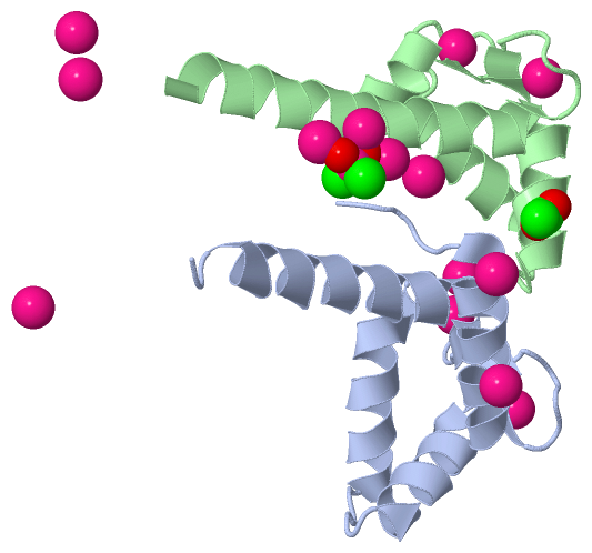
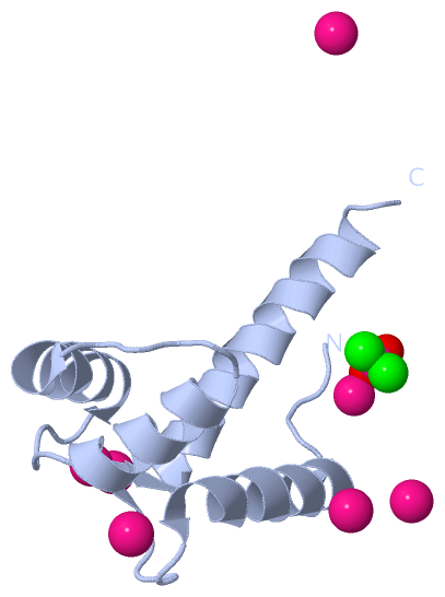
Ligands, Modified Residues, Ions (3, 16)
Asymmetric Unit (3, 16)
|
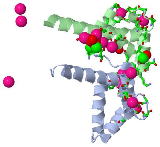
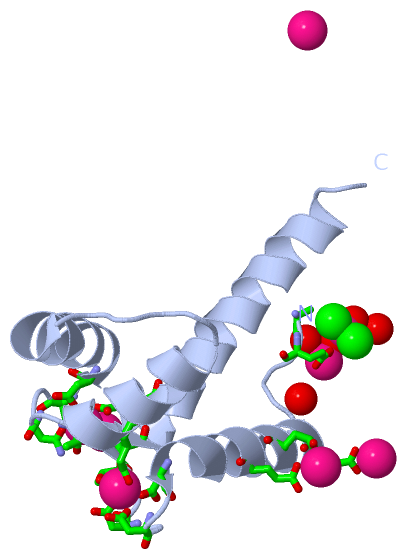
Sites (13, 13)
Asymmetric Unit (13, 13)
|
SS Bonds (0, 0)| (no "SS Bond" information available for 1N0Y) |
Cis Peptide Bonds (0, 0)| (no "Cis Peptide Bond" information available for 1N0Y) |
SAPs(SNPs)/Variants (0, 0)| (no "SAP(SNP)/Variant" information available for 1N0Y) |
PROSITE Motifs (2, 9)
Asymmetric Unit (2, 9)
|
||||||||||||||||||||||||||||||||||||||||||||||||||||||||||||||||||||||||||||||||||||||||||||||||
Exons (0, 0)| (no "Exon" information available for 1N0Y) |
Sequences/Alignments
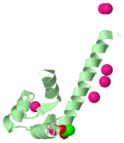
Asymmetric UnitChain A from PDB Type:PROTEIN Length:87 aligned with CALM_PARTE | P07463 from UniProtKB/Swiss-Prot Length:149 Alignment length:87 11 21 31 41 51 61 71 81 CALM_PARTE 2 AEQLTEEQIAEFKEAFALFDKDGDGTITTKELGTVMRSLGQNPTEAELQDMINEVDADGNGTIDFPEFLSLMARKMKEQDSEEELIE 88 SCOP domains d1n0ya_ A: Calmodulin SCOP domains CATH domains 1n0yA00 A:1-87 EF-hand CATH domains Pfam domains --------------------------------------------------------------------------------------- Pfam domains SAPs(SNPs) --------------------------------------------------------------------------------------- SAPs(SNPs) PROSITE (1) ------EF_HAND_2 PDB: A:7-42 UniProt: 8-43EF_HAND_2 PDB: A:43-78 -EF_HAND_ PROSITE (1) PROSITE (2) -------------------EF_HAND_1 -----------------------EF_HAND_1 ------------------- PROSITE (2) Transcript --------------------------------------------------------------------------------------- Transcript 1n0y A 1 AEQLTEEQIAEFKEAFALFDKDGDGTITTKELGTVMRSLGQNPTEAELQDMINEVDADGNGTIDFPEFLSLMARKMKEQDSEEELIE 87 10 20 30 40 50 60 70 80 Chain B from PDB Type:PROTEIN Length:79 aligned with CALM_PARTE | P07463 from UniProtKB/Swiss-Prot Length:149 Alignment length:79 79 89 99 109 119 129 139 CALM_PARTE 70 LSLMARKMKEQDSEEELIEAFKVFDRDGNGLISAAELRHVMTNLGEKLTDDEVDEMIREADIDGDGHINYEEFVRMMVS 148 SCOP domains d1n0yb_ B: Calmodulin SCOP domains CATH domains 1n0yB00 B:69-147 EF-hand CATH domains Pfam domains (1) ----------EF_hand_5-1n0yB01 B:79-145 -- Pfam domains (1) Pfam domains (2) ----------EF_hand_5-1n0yB02 B:79-145 -- Pfam domains (2) Pfam domains (3) ----------EF_hand_5-1n0yB03 B:79-145 -- Pfam domains (3) Pfam domains (4) ----------EF_hand_5-1n0yB04 B:79-145 -- Pfam domains (4) SAPs(SNPs) ------------------------------------------------------------------------------- SAPs(SNPs) PROSITE (1) EF_HAND_2 -EF_HAND_2 PDB: B:80-115 EF_HAND_2 PDB: B:116-147 PROSITE (1) PROSITE (2) ------------------------EF_HAND_1 -----------------------EF_HAND_1 ------ PROSITE (2) Transcript ------------------------------------------------------------------------------- Transcript 1n0y B 69 LSLMARKMKEQDSEEELIEAFKVFDRDGNGLISAAELRHVMTNLGEKLTDDEVDEMIREADIDGDGHINYEEFVRMMVS 147 78 88 98 108 118 128 138
|
||||||||||||||||||||
SCOP Domains (1, 2)
Asymmetric Unit
 |
CATH Domains (1, 2)
Asymmetric Unit
|
Pfam Domains (1, 4)
Asymmetric Unit
|
Gene Ontology (2, 2)|
Asymmetric Unit(hide GO term definitions) Chain A,B (CALM_PARTE | P07463)
|
||||||||||||||||||
Interactive Views
|
|||||||||||||||||||||||||||||||||||||||||||||||||||||||||||||||||||||||||||||||||||||||||||||||||||||||||||||||||||||||||||||||||||||||||||||||||||||||||||||||||||||||||||||||||||||||||||||||||||||||||||||||||||||||||||||||||||||||||||||||
Still Images
|
||||||||||||||||
Databases
|
||||||||||||||||||||||||||||||||||||||||||||||||||||||||||||||||||||||||||||||||||||||||||||||||||||||||||||||||||||||||||||||||||||||||||||||||||||||||||||||||
Analysis Tools
|
|||||||||||||||||||||||||||||||||||||||||||||||||||||||||||||
Entries Sharing at Least One Protein Chain (UniProt ID)
Related Entries Specified in the PDB File
|
|