|
|
|
|
 Description
|
|
Compounds
|
||||||||||||||||||||||||||||||||||||||||||||||||||||||||||||||||||
Chains, Units
Summary Information (see also Sequences/Alignments below) |
Ligands, Modified Residues, Ions (2, 7)| Asymmetric/Biological Unit (2, 7) |
Sites (2, 2)
Asymmetric Unit (2, 2)
|
SS Bonds (0, 0)| (no "SS Bond" information available for 1MZP) |
Cis Peptide Bonds (0, 0)| (no "Cis Peptide Bond" information available for 1MZP) |
SAPs(SNPs)/Variants (0, 0)| (no "SAP(SNP)/Variant" information available for 1MZP) |
PROSITE Motifs (1, 1)
Asymmetric/Biological Unit (1, 1)
|
||||||||||||||||||||||||
Exons (0, 0)| (no "Exon" information available for 1MZP) |
Sequences/Alignments
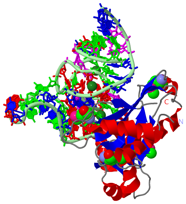
Asymmetric/Biological UnitChain A from PDB Type:PROTEIN Length:217 aligned with RL1_SULAC | P35024 from UniProtKB/Swiss-Prot Length:221 Alignment length:217 13 23 33 43 53 63 73 83 93 103 113 123 133 143 153 163 173 183 193 203 213 RL1_SULAC 4 VLADKESLIEALKLALSTEYNVKRNFTQSVEIILTFKGIDMKKGDLKLREIVPLPKQPSKAKRVLVVPSFEQLEYAKKASPNVVITREELQKLQGQKRPVKKLARQNEWFLINQESMALAGRILGPALGPRGKFPTPLPNTADISEYINRFKRSVLVKTKDQPQVQVFIGTEDMKPEDLAENAIAVLNAIENKAKVETNLRNIYVKTTMGKAVKVKR 220 SCOP domains d1mzpa_ A: Ribosomal protein L1 SCOP domains CATH domains 1mzpA01 A:1-61,A:153-217 [code=3.30.190.20, no name defined]1mzpA02 A:62-152 [code=3.40.50.790, no name defined] 1mzpA01 A:1-61,A:153-217 [code=3.30.190.20, no name defined] CATH domains Pfam domains --Ribosomal_L1-1mzpA01 A:3-212 ----- Pfam domains SAPs(SNPs) ------------------------------------------------------------------------------------------------------------------------------------------------------------------------------------------------------------------------- SAPs(SNPs) PROSITE --------------------------------------------------------------------------------------------------------------------RIBOSOMAL_L1 --------------------------------------------------------------------------------- PROSITE Transcript ------------------------------------------------------------------------------------------------------------------------------------------------------------------------------------------------------------------------- Transcript 1mzp A 1 MLADKESLIEALKLALSTEYNVKRNFTQSVEIILTFKGIDmKKGDLKLREIVPLPKQPSKAKRVLVVPSSEQLEYAKKASPKVVITREELQKLQGQKRPVKKLARQNEWFLINQESmALAGRILGPALGPRGKFPTPLPNTADISEYINRFKRSVLVKTKDQPQVQVFIGTEDmKPEDLAENAIAVLNAIENKAKVETNLRNIYVKTTmGKAVKVKR 217 10 20 30 40| 50 60 70 80 90 100 110 |120 130 140 150 160 170 | 180 190 200 210 41-MSE 117-MSE 174-MSE 209-MSE
Chain B from PDB  Type:RNA  Length:55

                 1mzp B   1 GGGAUGCGUAGGAUAGGUGGGAGCCGCAAGGCGCCGGUGAAAUACCACCCUUCCC  55
                                    10        20        30        40        50
|
||||||||||||||||||||
SCOP Domains (1, 1)
Asymmetric/Biological Unit
 |
CATH Domains (2, 2)
Asymmetric/Biological Unit
|
Pfam Domains (1, 1)
Asymmetric/Biological Unit
|
Gene Ontology (9, 9)|
Asymmetric/Biological Unit(hide GO term definitions) Chain A (RL1_SULAC | P35024)
|
||||||||||||||||||||||||||||||||||||||||||||||||||||||||||||||||||||||||
Interactive Views
|
||||||||||||||||||||||||||||||||||||||||||||||||||||||||||||||||||||||||||||||||||||||||||||||||||||||||||||||||||||||||||||||||||||
Still Images
|
||||||||||||||||
Databases
|
||||||||||||||||||||||||||||||||||||||||||||||||||||||||||||||||||||||||||||||||||||||||||||||||||||||||||||||||||||||||||||||||||||||||||||||||||||||||||||||||
Analysis Tools
|
|||||||||||||||||||||||||||||||||||||||||||||||||||||||||||||
Entries Sharing at Least One Protein Chain (UniProt ID)
Related Entries Specified in the PDB File
|
|