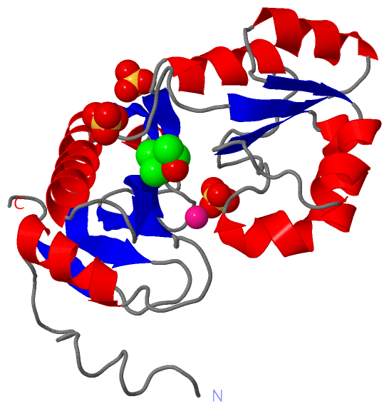
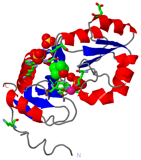
Show PDB file:   
         Plain Text   HTML   (compressed file size)
QuickSearch:   
by PDB,NDB,UniProt,PROSITE Code or Search Term(s)  
(-)Asym./Biol. Unit - manually
(-)Asym./Biol. Unit
(-)Asym./Biol. Unit - sites
collapse expand < >
Image Asym./Biol. Unit - manually
Asym./Biol. Unit - manually  (Jmol Viewer)
Image Asym./Biol. Unit
Asym./Biol. Unit  (Jmol Viewer)
Image Asym./Biol. Unit - sites
Asym./Biol. Unit - sites  (Jmol Viewer)

(-) Description

Title :  RIBOSOMAL PROTEIN L1 MUTANT WITH SERINE 179 REPLACED BY CYSTEINE
 
Authors :  J. Unge, S. Al-Karadaghi, A. Liljas, B. -H. Jonsson, I. Eliseikina, N. Ossina, N. Nevskaya, N. Fomenkova, M. Garber, S. Nikonov
Date :  20 Feb 97  (Deposition) - 15 May 97  (Release) - 24 Feb 09  (Revision)
Method :  X-RAY DIFFRACTION
Resolution :  1.90
Chains :  Asym./Biol. Unit :  A
Keywords :  Ribosomal Protein, Rna Binding, Protein Synthesis, Mutant (Keyword Search: [Gene Ontology, PubMed, Web (Google))
 
Reference :  J. Unge, S. Al-Karadaghi, A. Liljas, B. H. Jonsson, I. Eliseikina, N. Ossina, N. Nevskaya, N. Fomenkova, M. Garber, S. Nikonov
A Mutant Form Of The Ribosomal Protein L1 Reveals Conformational Flexibility.
Febs Lett. V. 411 53 1997
PubMed-ID: 9247141  |  Reference-DOI: 10.1016/S0014-5793(97)00611-X
(for further references see the PDB file header)

(-) Compounds

Molecule 1 - RIBOSOMAL PROTEIN L1
    ChainsA
    EngineeredYES
    Expression SystemESCHERICHIA COLI BL21
    Expression System PlasmidPACA-L1
    Expression System StrainBL21
    Expression System Taxid511693
    MutationYES
    OrganelleRIBOSOME
    Organism ScientificTHERMUS THERMOPHILUS
    Organism Taxid274
    StrainVK-1
    SynonymTL2

 Structural Features

(-) Chains, Units

  1
Asymmetric/Biological Unit A

Summary Information (see also Sequences/Alignments below)

(-) Ligands, Modified Residues, Ions  (3, 5)

Asymmetric/Biological Unit (3, 5)
No.NameCountTypeFull Name
1HG1Ligand/IonMERCURY (II) ION
2MRD1Ligand/Ion(4R)-2-METHYLPENTANE-2,4-DIOL
3SO43Ligand/IonSULFATE ION

(-) Sites  (5, 5)

Asymmetric Unit (5, 5)
No.NameEvidenceResiduesDescription
1AC1SOFTWARELYS A:18 , THR A:61 , HIS A:199BINDING SITE FOR RESIDUE SO4 A 800
2AC2SOFTWAREHIS A:66 , GLY A:67 , GLN A:71 , LYS A:184BINDING SITE FOR RESIDUE SO4 A 801
3AC3SOFTWAREGLU A:88 , LYS A:141 , ASP A:166 , LYS A:167 , HOH A:683BINDING SITE FOR RESIDUE SO4 A 803
4AC4SOFTWAREGLU A:42 , HIS A:172BINDING SITE FOR RESIDUE HG A 900
5AC5SOFTWAREGLY A:67 , GLY A:176 , LYS A:177BINDING SITE FOR RESIDUE MRD A 700

(-) SS Bonds  (0, 0)

(no "SS Bond" information available for 1AD2)

(-) Cis Peptide Bonds  (0, 0)

(no "Cis Peptide Bond" information available for 1AD2)

 Sequence-Structure Mapping

(-) SAPs(SNPs)/Variants  (0, 0)

(no "SAP(SNP)/Variant" information available for 1AD2)

(-) PROSITE Motifs  (1, 1)

Asymmetric/Biological Unit (1, 1)
 PROSITEUniProtKBPDB
No.IDACDescriptionIDLocationCountLocation
1RIBOSOMAL_L1PS01199 Ribosomal protein L1 signature.RL1_THETH121-140  1A:120-139

(-) Exons   (0, 0)

(no "Exon" information available for 1AD2)

(-) Sequences/Alignments

Asymmetric/Biological Unit
   Reformat: Number of residues per line =  ('0' or empty: single-line sequence representation)
  Number of residues per labelling interval =   
  UniProt sequence: complete  aligned part    
   Show mapping: SCOP domains CATH domains Pfam domains Secondary structure (by author)
SAPs(SNPs) PROSITE motifs Exons
(details for a mapped element are shown in a popup box when the mouse pointer rests over it)
Chain A from PDB  Type:PROTEIN  Length:224
 aligned with RL1_THETH | P27150 from UniProtKB/Swiss-Prot  Length:229

    Alignment length:224
                                    15        25        35        45        55        65        75        85        95       105       115       125       135       145       155       165       175       185       195       205       215       225    
            RL1_THETH     6 KRYRALLEKVDPNKIYTIDEAAHLVKELATAKFDETVEVHAKLGIDPRRSDQNVRGTVSLPHGLGKQVRVLAIAKGEKIKEAEEAGADYVGGEEIIQKILDGWMDFDAVVATPDVMGAVGSKLGRILGPRGLLPNPKAGTVGFNIGEIIREIKAGRIEFRNDKTGAIHAPVGKASFPPEKLADNIRAFIRALEAHKPEGAKGTFLRSVYVTTTMGPSVRINPHS 229
               SCOP domains d1ad2a_ A: Ribosomal protein L1                                                                                                                                                                                                  SCOP domains
               CATH domains ----------1ad2A01 A:15-66,A:160-228                           1ad2A02 A:67-159  [code=3.40.50.790, no name defined]                                        1ad2A01 A:15-66,A:160-228  [code=3.30.190.20, no name defined]        CATH domains
               Pfam domains -------------------------------------------------------------------------------------------------------------------------------------------------------------------------------------------------------------------------------- Pfam domains
         Sec.struct. author .................hhhhhhhhhh........eeeeeeee......hhh..eeeee..........eeee..hhhhhhhhh....eee.hhhhhhhh.......eeee.hhhhhhhhhhhhhhhhhh..............hhhhhhhhh..eeeee.....eeeeeeee....hhhhhhhhhhhhhhhhhh.........eeeeeee......eee.... Sec.struct. author
                 SAPs(SNPs) -------------------------------------------------------------------------------------------------------------------------------------------------------------------------------------------------------------------------------- SAPs(SNPs)
                    PROSITE -------------------------------------------------------------------------------------------------------------------RIBOSOMAL_L1        ----------------------------------------------------------------------------------------- PROSITE
                 Transcript -------------------------------------------------------------------------------------------------------------------------------------------------------------------------------------------------------------------------------- Transcript
                 1ad2 A   5 KRYRALLEKVDPNKIYTIDEAAHLVKELATAKFDETVEVHAKLGIDPRRSDQNVRGTVSLPHGLGKQVRVLAIAKGEKIKEAEEAGADYVGGEEIIQKILDGWMDFDAVVATPDVMGAVGSKLGRILGPRGLLPNPKAGTVGFNIGEIIREIKAGRIEFRNDKTGAIHAPVGKACFPPEKLADNIRAFIRALEAHKPEGAKGTFLRSVYVTTTMGPSVRINPHS 228
                                    14        24        34        44        54        64        74        84        94       104       114       124       134       144       154       164       174       184       194       204       214       224    

   Legend:   → Mismatch (orange background)
  - → Gap (green background, '-', border residues have a numbering label)
    → Modified Residue (blue background, lower-case, 'x' indicates undefined single-letter code, labelled with number + name)
  x → Chemical Group (purple background, 'x', labelled with number + name, e.g. ACE or NH2)
  extra numbering lines below/above indicate numbering irregularities and modified residue names etc., number ends below/above '|'

 Classification and Annotation

(-) SCOP Domains  (1, 1)

Asymmetric/Biological Unit

(-) CATH Domains  (2, 2)

Asymmetric/Biological Unit
(-)
Class: Alpha Beta (26913)

(-) Pfam Domains  (0, 0)

(no "Pfam Domain" information available for 1AD2)

(-) Gene Ontology  (9, 9)

Asymmetric/Biological Unit(hide GO term definitions)
Chain A   (RL1_THETH | P27150)
molecular function
    GO:0003723    RNA binding    Interacting selectively and non-covalently with an RNA molecule or a portion thereof.
    GO:0019843    rRNA binding    Interacting selectively and non-covalently with ribosomal RNA.
    GO:0003735    structural constituent of ribosome    The action of a molecule that contributes to the structural integrity of the ribosome.
    GO:0000049    tRNA binding    Interacting selectively and non-covalently with transfer RNA.
biological process
    GO:0006417    regulation of translation    Any process that modulates the frequency, rate or extent of the chemical reactions and pathways resulting in the formation of proteins by the translation of mRNA or circRNA.
    GO:0006412    translation    The cellular metabolic process in which a protein is formed, using the sequence of a mature mRNA or circRNA molecule to specify the sequence of amino acids in a polypeptide chain. Translation is mediated by the ribosome, and begins with the formation of a ternary complex between aminoacylated initiator methionine tRNA, GTP, and initiation factor 2, which subsequently associates with the small subunit of the ribosome and an mRNA or circRNA. Translation ends with the release of a polypeptide chain from the ribosome.
cellular component
    GO:0030529    intracellular ribonucleoprotein complex    An intracellular macromolecular complex containing both protein and RNA molecules.
    GO:0015934    large ribosomal subunit    The larger of the two subunits of a ribosome. Two sites on the ribosomal large subunit are involved in translation, namely the aminoacyl site (A site) and peptidyl site (P site).
    GO:0005840    ribosome    An intracellular organelle, about 200 A in diameter, consisting of RNA and protein. It is the site of protein biosynthesis resulting from translation of messenger RNA (mRNA). It consists of two subunits, one large and one small, each containing only protein and RNA. Both the ribosome and its subunits are characterized by their sedimentation coefficients, expressed in Svedberg units (symbol: S). Hence, the prokaryotic ribosome (70S) comprises a large (50S) subunit and a small (30S) subunit, while the eukaryotic ribosome (80S) comprises a large (60S) subunit and a small (40S) subunit. Two sites on the ribosomal large subunit are involved in translation, namely the aminoacyl site (A site) and peptidyl site (P site). Ribosomes from prokaryotes, eukaryotes, mitochondria, and chloroplasts have characteristically distinct ribosomal proteins.

 Visualization

(-) Interactive Views

Asymmetric/Biological Unit
  Complete Structure
    Jena3D(integrated viewing of ligand, site, SAP, PROSITE, SCOP information)
    WebMol | AstexViewer[tm]@PDBe
(Java Applets, require no local installation except for Java; loading may be slow)
    STRAP
(Java WebStart application, automatic local installation, requires Java; full application with system access!)
    RasMol
(require local installation)
    Molscript (VRML)
(requires installation of a VRML viewer; select preferred view via VRML and generate a mono or stereo PDF format file)
 
  Ligands, Modified Residues, Ions
    HG  [ RasMol | Jena3D ]  +environment [ RasMol | Jena3D ]
    MRD  [ RasMol | Jena3D ]  +environment [ RasMol | Jena3D ]
    SO4  [ RasMol | Jena3D ]  +environment [ RasMol | Jena3D ]
 
  Sites
    AC1  [ RasMol ]  +environment [ RasMol ]
    AC2  [ RasMol ]  +environment [ RasMol ]
    AC3  [ RasMol ]  +environment [ RasMol ]
    AC4  [ RasMol ]  +environment [ RasMol ]
    AC5  [ RasMol ]  +environment [ RasMol ]
 
  Cis Peptide Bonds
(no "Cis Peptide Bonds" information available for 1ad2)
 

(-) Still Images

Jmol
  protein: cartoon or spacefill or dots and stick; nucleic acid: cartoon and stick; ligands: spacefill; active site: stick
Molscript
  protein, nucleic acid: cartoon; ligands: spacefill; active site: ball and stick

 Databases and Analysis Tools

(-) Databases

Access by PDB/NDB ID
  1ad2
    Family and Domain InformationProDom | SYSTERS
    General Structural InformationGlycoscienceDB | MMDB | NDB | OCA | PDB | PDBe | PDBj | PDBsum | PDBWiki | PQS | PROTEOPEDIA
    Orientation in MembranesOPM
    Protein SurfaceSURFACE
    Secondary StructureDSSP (structure derived) | HSSP (homology derived)
    Structural GenomicsGeneCensus
    Structural NeighboursCE | VAST
    Structure ClassificationCATH | Dali | SCOP
    Validation and Original DataBMRB Data View | BMRB Restraints Grid | EDS | PROCHECK | RECOORD | WHAT_CHECK
 
Access by UniProt ID/Accession number
  RL1_THETH | P27150
    Comparative Protein Structure ModelsModBase
    Genomic InformationEnsembl
    Protein-protein InteractionDIP
    Sequence, Family and Domain InformationInterPro | Pfam | SMART | UniProtKB/SwissProt
 
Access by Enzyme Classificator   (EC Number)
  (no 'Enzyme Classificator' available)
    General Enzyme InformationBRENDA | EC-PDB | Enzyme | IntEnz
    PathwayKEGG | MetaCyc
 
Access by Disease Identifier   (MIM ID)
  (no 'MIM ID' available)
    Disease InformationOMIM
 
Access by GenAge ID
  (no 'GenAge ID' available)
    Age Related InformationGenAge

(-) Analysis Tools

Access by PDB/NDB ID
    Domain InformationXDom
    Interatomic Contacts of Structural UnitsCSU
    Ligand-protein ContactsLPC
    Protein CavitiescastP
    Sequence and Secondary StructurePDBCartoon
    Structure AlignmentSTRAP(Java WebStart application, automatic local installation, requires Java; full application with system access!)
    Structure and Sequence BrowserSTING
 
Access by UniProt ID/Accession number
  RL1_THETH | P27150
    Protein Disorder PredictionDisEMBL | FoldIndex | GLOBPLOT (for more information see DisProt)

 Related Entries

(-) Entries Sharing at Least One Protein Chain (UniProt ID)

UniProtKB/Swiss-Prot
        RL1_THETH | P271501eg0 1zho 2hw8 2oum 2ov7 2vpl 3tg8 3u4m 3u56 3umy 487d 4f9t 4qg3 4qgb 4qvi 4reo 5ib7 5ib8 5ibb 5imq 5imr 5j8b

(-) Related Entries Specified in the PDB File

(no "Related Entries Specified in the PDB File" available for 1AD2)