| molecular function |
|---|
| | GO:0004871 | | signal transducer activity | | Conveys a signal across a cell to trigger a change in cell function or state. A signal is a physical entity or change in state that is used to transfer information in order to trigger a response. |
| biological process |
|---|
| | GO:0007186 | | G-protein coupled receptor signaling pathway | | A series of molecular signals that proceeds with an activated receptor promoting the exchange of GDP for GTP on the alpha-subunit of an associated heterotrimeric G-protein complex. The GTP-bound activated alpha-G-protein then dissociates from the beta- and gamma-subunits to further transmit the signal within the cell. The pathway begins with receptor-ligand interaction, or for basal GPCR signaling the pathway begins with the receptor activating its G protein in the absence of an agonist, and ends with regulation of a downstream cellular process, e.g. transcription. The pathway can start from the plasma membrane, Golgi or nuclear membrane (PMID:24568158 and PMID:16902576). |
| | GO:0042462 | | eye photoreceptor cell development | | Development of a photoreceptor, a sensory cell in the eye that reacts to the presence of light. They usually contain a pigment that undergoes a chemical change when light is absorbed, thus stimulating a nerve. |
| | GO:0007602 | | phototransduction | | The sequence of reactions within a cell required to convert absorbed photons into a molecular signal. |
| | GO:0008104 | | protein localization | | Any process in which a protein is transported to, or maintained in, a specific location. |
| | GO:0022400 | | regulation of rhodopsin mediated signaling pathway | | Any process that modulates the frequency, rate or extent of rhodopsin-mediated signaling. |
| | GO:0016056 | | rhodopsin mediated signaling pathway | | The series of molecular signals generated as a consequence of excitation of rhodopsin by a photon and the events that convert the absorbed photons into a cellular response. |
| | GO:0007165 | | signal transduction | | The cellular process in which a signal is conveyed to trigger a change in the activity or state of a cell. Signal transduction begins with reception of a signal (e.g. a ligand binding to a receptor or receptor activation by a stimulus such as light), or for signal transduction in the absence of ligand, signal-withdrawal or the activity of a constitutively active receptor. Signal transduction ends with regulation of a downstream cellular process, e.g. regulation of transcription or regulation of a metabolic process. Signal transduction covers signaling from receptors located on the surface of the cell and signaling via molecules located within the cell. For signaling between cells, signal transduction is restricted to events at and within the receiving cell. |
| cellular component |
|---|
| | GO:0005834 | | heterotrimeric G-protein complex | | Any of a family of heterotrimeric GTP-binding and hydrolyzing proteins; they belong to a superfamily of GTPases that includes monomeric proteins such as EF-Tu and RAS. Heterotrimeric G-proteins consist of three subunits; the alpha subunit contains the guanine nucleotide binding site and possesses GTPase activity; the beta and gamma subunits are tightly associated and function as a beta-gamma heterodimer; extrinsic plasma membrane proteins (cytoplasmic face) that function as a complex to transduce signals from G-protein coupled receptors to an effector protein. |
| | GO:0016020 | | membrane | | A lipid bilayer along with all the proteins and protein complexes embedded in it an attached to it. |
| | GO:0097381 | | photoreceptor disc membrane | | Ovally-shaped membranous stack located inside the photoreceptor outer segment, and containing densely packed molecules of the photoreceptor protein rhodopsin that traverse the lipid bilayer. Disc membranes are apparently derived from the plasma membrane in the region of the cilium that connects the photoreceptor outer segment to the inner segment. |
| | GO:0005886 | | plasma membrane | | The membrane surrounding a cell that separates the cell from its external environment. It consists of a phospholipid bilayer and associated proteins. |
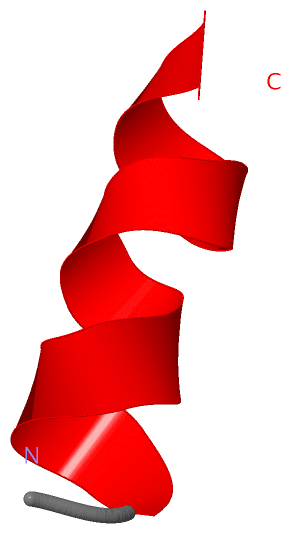
 Description
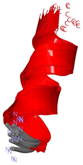