Show PDB file:   
         Plain Text   HTML   (compressed file size)
QuickSearch:   
by PDB,NDB,UniProt,PROSITE Code or Search Term(s)  
(-)Asym./Biol. Unit
(-)Asym./Biol. Unit - sites
collapse expand < >
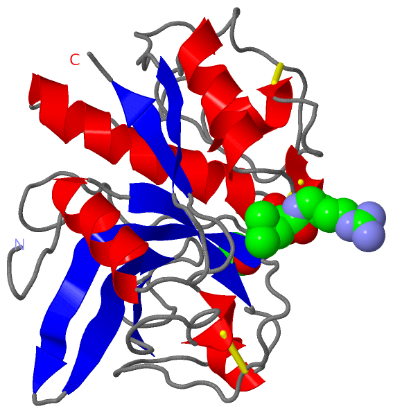
Image Asym./Biol. Unit
Asym./Biol. Unit  (Jmol Viewer)
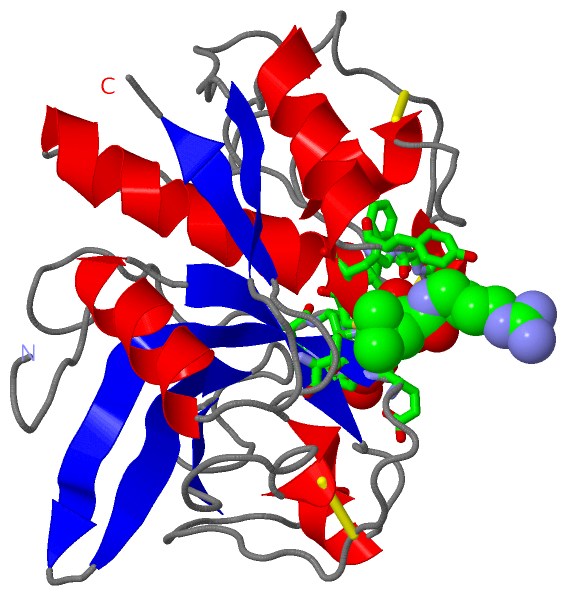
Image Asym./Biol. Unit - sites
Asym./Biol. Unit - sites  (Jmol Viewer)

(-) Description

Title :  CRYSTAL STRUCTURE OF A CARICAIN D158E MUTANT IN COMPLEX WITH E-64
 
Authors :  N. A. Katerelos
Date :  04 May 96  (Deposition) - 11 Jan 97  (Release) - 24 Feb 09  (Revision)
Method :  X-RAY DIFFRACTION
Resolution :  2.00
Chains :  Asym./Biol. Unit :  A
Keywords :  Cysteine Proteinase, Thiol Protease, Hydrolase (Keyword Search: [Gene Ontology, PubMed, Web (Google)] )
 
Reference :  N. A. Katerelos, M. A. Taylor, M. Scott, P. W. Goodenough, R. W. Pickersgill
Crystal Structure Of A Caricain D158E Mutant In Complex With E-64.
Febs Lett. V. 392 35 1996
PubMed-ID: 8769310  |  Reference-DOI: 10.1016/0014-5793(96)00697-7
(for further references see the PDB file header)

(-) Compounds

Molecule 1 - CARICAIN
    Chains: A
    EC Number: 3.4.22.30
    Engineered: YES
    Expression System: ESCHERICHIA COLI
    Expression System Plasmid: PLYSS
    Expression System Strain: PET
    Expression System Taxid: 562
    Expression System Vector: PET VECTOR
    Expression System Vector Type: PET
    Mutation: YES
    Organism Common: PAPAYA
    Organism Scientific: CARICA PAPAYA
    Organism Taxid: 3649
    Synonym: PAPAYA PROTEINASE, PROTEASE OMEGA

 Structural Features

(-) Chains, Units

  1
Asymmetric/Biological Unit : A

Summary Information (see also Sequences/Alignments below)

(-) Ligands, Modified Residues, Ions  (2, 2)

Asymmetric/Biological Unit (2, 2)
No.NameCountTypeFull Name
1E641Ligand/IonN-[N-[1-HYDROXYCARBOXYETHYL-CARBONYL]LEUCYLAMINO-BUTYL]-GUANIDINE
2EOH1Ligand/IonETHANOL

(-) Sites  (3, 3)

Asymmetric Unit (3, 3)
No.NameEvidenceResiduesDescription
1AC1SOFTWAREGLN A:19 , GLY A:23 , SER A:24 , CYS A:25 , TRP A:26 , GLY A:65 , GLY A:66 , TYR A:67 , VAL A:133 , GLU A:158 , HIS A:159 , HOH A:310BINDING SITE FOR RESIDUE E64 A 217
2AC2SOFTWAREHIS A:18 , GLY A:20 , ALA A:90 , HOH A:312BINDING SITE FOR RESIDUE EOH A 218
3ACTUNKNOWNCYS A:25 , HIS A:159ACTIVE SITE.

(-) SS Bonds  (3, 3)

Asymmetric/Biological Unit
No.Residues
1A:22 -A:63
2A:56 -A:95
3A:153 -A:204

(-) Cis Peptide Bonds  (1, 1)

Asymmetric/Biological Unit
No.Residues
1Gly A:151 -Pro A:152

 Sequence-Structure Mapping

(-) SAPs(SNPs)/Variants  (0, 0)

(no "SAP(SNP)/Variant" information available for 1MEG)

(-) PROSITE Motifs  (3, 3)

Asymmetric/Biological Unit (3, 3)
 PROSITEUniProtKBPDB
No.IDACDescriptionIDLocationCountLocation
1THIOL_PROTEASE_CYSPS00139 Eukaryotic thiol (cysteine) proteases cysteine active site.PAPA3_CARPA151-162  1A:19-30
2THIOL_PROTEASE_HISPS00639 Eukaryotic thiol (cysteine) proteases histidine active site.PAPA3_CARPA289-299  1A:157-167
3THIOL_PROTEASE_ASNPS00640 Eukaryotic thiol (cysteine) proteases asparagine active site.PAPA3_CARPA306-325  1A:174-193

(-) Exons   (0, 0)

(no "Exon" information available for 1MEG)

(-) Sequences/Alignments

Asymmetric/Biological Unit
   Reformat: Number of residues per line =  ('0' or empty: single-line sequence representation)
  Number of residues per labelling interval =   
  UniProt sequence: complete  aligned part    
   Show mapping: SCOP domains CATH domains Pfam domains Secondary structure (by author)
SAPs(SNPs) PROSITE motifs Exons
(details for a mapped element are shown in a popup box when the mouse pointer rests over it)
Chain A from PDB  Type:PROTEIN  Length:216
 aligned with PAPA3_CARPA | P10056 from UniProtKB/Swiss-Prot  Length:348

    Alignment length:216
                                   142       152       162       172       182       192       202       212       222       232       242       252       262       272       282       292       302       312       322       332       342      
          PAPA3_CARPA   133 LPENVDWRKKGAVTPVRHQGSCGSCWAFSAVATVEGINKIRTGKLVELSEQELVDCERRSHGCKGGYPPYALEYVAKNGIHLRSKYPYKAKQGTCRAKQVGGPIVKTSGVGRVQPNNEGNLLNAIAKQPVSVVVESKGRPFQLYKGGIFEGPCGTKVDHAVTAVGYGKSGGKGYILIKNSWGTAWGEKGYIRIKRAPGNSPGVCGLYKSSYYPTKN 348
               SCOP domains d1mega_ A: Caricain (protease omega)                                                                                                                                                                                     SCOP domains
               CATH domains 1megA00 A:1-216 Cysteine proteinases                                                                                                                                                                                     CATH domains
               Pfam domains Peptidase_C1-1megA01 A:1-214                                                                                                                                                                                          -- Pfam domains
         Sec.struct. author ........................hhhhhhhhhhhhhhhhhh.......hhhhhhh.....hhh...hhhhhhhhhhh..............................eeee.....hhhhhhhhhh..eeeee....hhhh................eeeeeeeeeee..eeeeeee...........eeeee........hhh.....eeee.. Sec.struct. author
                 SAPs(SNPs) ------------------------------------------------------------------------------------------------------------------------------------------------------------------------------------------------------------------------ SAPs(SNPs)
                    PROSITE ------------------THIOL_PROTEA------------------------------------------------------------------------------------------------------------------------------THIOL_PROTE------THIOL_PROTEASE_ASN  ----------------------- PROSITE
                 Transcript ------------------------------------------------------------------------------------------------------------------------------------------------------------------------------------------------------------------------ Transcript
                 1meg A   1 LPENVDWRKKGAVTPVRHQGSCGSCWAFSAVATVEGINKIRTGKLVELSEQELVDCERRSHGCKGGYPPYALEYVAKNGIHLRSKYPYKAKQGTCRAKQVGGPIVKTSGVGRVQPNNEGNLLNAIAKQPVSVVVESKGRPFQLYKGGIFEGPCGTKVEHAVTAVGYGKSGGKGYILIKNSWGTAWGEKGYIRIKRAPGNSPGVCGLYKSSYYPTKN 216
                                    10        20        30        40        50        60        70        80        90       100       110       120       130       140       150       160       170       180       190       200       210      

   Legend:   → Mismatch (orange background)
  - → Gap (green background, '-', border residues have a numbering label)
    → Modified Residue (blue background, lower-case, 'x' indicates undefined single-letter code, labelled with number + name)
  x → Chemical Group (purple background, 'x', labelled with number + name, e.g. ACE or NH2)
  extra numbering lines below/above indicate numbering irregularities and modified residue names etc., number ends below/above '|'

 Classification and Annotation

(-) SCOP Domains  (1, 1)

Asymmetric/Biological Unit

(-) CATH Domains  (1, 1)

Asymmetric/Biological Unit
(-)
Class: Alpha Beta (26913)

(-) Pfam Domains  (1, 1)

Asymmetric/Biological Unit

(-) Gene Ontology  (4, 4)

Asymmetric/Biological Unit(hide GO term definitions)
Chain A   (PAPA3_CARPA | P10056)
molecular function
    GO:0008234    cysteine-type peptidase activity    Catalysis of the hydrolysis of peptide bonds in a polypeptide chain by a mechanism in which the sulfhydryl group of a cysteine residue at the active center acts as a nucleophile.
    GO:0016787    hydrolase activity    Catalysis of the hydrolysis of various bonds, e.g. C-O, C-N, C-C, phosphoric anhydride bonds, etc. Hydrolase is the systematic name for any enzyme of EC class 3.
    GO:0008233    peptidase activity    Catalysis of the hydrolysis of a peptide bond. A peptide bond is a covalent bond formed when the carbon atom from the carboxyl group of one amino acid shares electrons with the nitrogen atom from the amino group of a second amino acid.
biological process
    GO:0006508    proteolysis    The hydrolysis of proteins into smaller polypeptides and/or amino acids by cleavage of their peptide bonds.

 Visualization

(-) Interactive Views

Asymmetric/Biological Unit
  Complete Structure
    Jena3D(integrated viewing of ligand, site, SAP, PROSITE, SCOP information)
    WebMol | AstexViewer[tm]@PDBe
(Java Applets, require no local installation except for Java; loading may be slow)
    STRAP
(Java WebStart application, automatic local installation, requires Java; full application with system access!)
    RasMol
(require local installation)
    Molscript (VRML)
(requires installation of a VRML viewer; select preferred view via VRML and generate a mono or stereo PDF format file)
 
  Ligands, Modified Residues, Ions
    E64  [ RasMol | Jena3D ]  +environment [ RasMol | Jena3D ]
    EOH  [ RasMol | Jena3D ]  +environment [ RasMol | Jena3D ]
 
  Sites
    AC1  [ RasMol ]  +environment [ RasMol ]
    AC2  [ RasMol ]  +environment [ RasMol ]
    ACT  [ RasMol ]  +environment [ RasMol ]
 
  Cis Peptide Bonds
    Gly A:151 - Pro A:152   [ RasMol ]  
 

(-) Still Images

Jmol
  protein: cartoon or spacefill or dots and stick; nucleic acid: cartoon and stick; ligands: spacefill; active site: stick
Molscript
  protein, nucleic acid: cartoon; ligands: spacefill; active site: ball and stick

 Databases and Analysis Tools

(-) Databases

Access by PDB/NDB ID
  1meg
    Family and Domain Information: ProDom | SYSTERS
    General Structural Information: GlycoscienceDB | MMDB | NDB | OCA | PDB | PDBe | PDBj | PDBsum | PDBWiki | PQS | PROTEOPEDIA
    Orientation in Membranes: OPM
    Protein Surface: SURFACE
    Secondary Structure: DSSP (structure derived) | HSSP (homology derived)
    Structural Genomics: GeneCensus
    Structural Neighbours: CE | VAST
    Structure Classification: CATH | Dali | SCOP
    Validation and Original Data: BMRB Data View | BMRB Restraints Grid | EDS | PROCHECK | RECOORD | WHAT_CHECK
 
Access by UniProt ID/Accession number
  PAPA3_CARPA | P10056
    Comparative Protein Structure Models: ModBase
    Genomic Information: Ensembl
    Protein-protein Interaction: DIP
    Sequence, Family and Domain Information: InterPro | Pfam | SMART | UniProtKB/SwissProt
 
Access by Enzyme Classificator   (EC Number)
  3.4.22.30
    General Enzyme Information: BRENDA | EC-PDB | Enzyme | IntEnz
    Pathway: KEGG | MetaCyc
 
Access by Disease Identifier   (MIM ID)
  (no 'MIM ID' available)
    Disease Information: OMIM
 
Access by GenAge ID
  (no 'GenAge ID' available)
    Age Related Information: GenAge

(-) Analysis Tools

Access by PDB/NDB ID
    Domain Information: XDom
    Interatomic Contacts of Structural Units: CSU
    Ligand-protein Contacts: LPC
    Protein Cavities: castP
    Sequence and Secondary Structure: PDBCartoon
    Structure Alignment: STRAP(Java WebStart application, automatic local installation, requires Java; full application with system access!)
    Structure and Sequence Browser: STING
 
Access by UniProt ID/Accession number
  PAPA3_CARPA | P10056
    Protein Disorder Prediction: DisEMBL | FoldIndex | GLOBPLOT (for more information see DisProt)

 Related Entries

(-) Entries Sharing at Least One Protein Chain (UniProt ID)

UniProtKB/Swiss-Prot
        PAPA3_CARPA | P10056: 1c7x 1eux 1f1i 1pci 1ppo

(-) Related Entries Specified in the PDB File

(no "Related Entries Specified in the PDB File" available for 1MEG)