Show PDB file:   
         Plain Text   HTML   (compressed file size)
QuickSearch:   
by PDB,NDB,UniProt,PROSITE Code or Search Term(s)  
(-)Asym.Unit - manually
(-)Asymmetric Unit
(-)Asym. Unit - sites
(-)Biological Unit 1
(-)Biol. Unit 1 - sites
(-)Biological Unit 2
collapse expand < >
Image Asym.Unit - manually
Asym.Unit - manually  (Jmol Viewer)
Image Asymmetric Unit
Asymmetric Unit  (Jmol Viewer)
Image Asym. Unit - sites
Asym. Unit - sites  (Jmol Viewer)
Image Biological Unit 1
Biological Unit 1  (Jmol Viewer)
Image Biol. Unit 1 - sites
Biol. Unit 1 - sites  (Jmol Viewer)
Image Biological Unit 2
Biological Unit 2  (Jmol Viewer)

(-) Description

Title :  CA2+-BINDING MIMICRY IN THE CRYSTAL STRUCTURE OF THE EU3+-BOUND MUTANT HUMAN MACROPHAGE CAPPING PROTEIN CAP G
 
Authors :  S. M. Vorobiev, F. S. Southwick, S. C. Almo
Date :  28 Jun 01  (Deposition) - 28 Jun 02  (Release) - 13 Jul 11  (Revision)
Method :  X-RAY DIFFRACTION
Resolution :  2.80
Chains :  Asym. Unit :  A
Biol. Unit 1:  A  (1x)
Biol. Unit 2:  A  (2x)
Keywords :  Actin, Human, Capping, Cap G, Macrophage, Gcap39, Mbhl, Gelsolin, Structural Protein (Keyword Search: [Gene Ontology, PubMed, Web (Google))
 
Reference :  S. M. Vorobiev, F. S. Southwick, S. C. Almo
Crystal Structure Of Mutant Human Macrophage Capping Protei
To Be Published
PubMed: search
(for further references see the PDB file header)

(-) Compounds

Molecule 1 - MACROPHAGE CAPPING PROTEIN
    ChainsA
    EngineeredYES
    Expression SystemESCHERICHIA COLI
    Expression System Taxid562
    MutationYES
    Organism CommonHUMAN
    Organism ScientificHOMO SAPIENS
    Organism Taxid9606
    SynonymACTIN-REGULATORY PROTEIN CAP-G

 Structural Features

(-) Chains, Units

  1
Asymmetric Unit A
Biological Unit 1 (1x)A
Biological Unit 2 (2x)A

Summary Information (see also Sequences/Alignments below)

(-) Ligands, Modified Residues, Ions  (1, 2)

Asymmetric Unit (1, 2)
No.NameCountTypeFull Name
1EU32Ligand/IonEUROPIUM (III) ION
Biological Unit 1 (1, 2)
No.NameCountTypeFull Name
1EU32Ligand/IonEUROPIUM (III) ION
Biological Unit 2 (1, 4)
No.NameCountTypeFull Name
1EU34Ligand/IonEUROPIUM (III) ION

(-) Sites  (2, 2)

Asymmetric Unit (2, 2)
No.NameEvidenceResiduesDescription
1AC1SOFTWAREGLY A:45 , ASP A:46 , GLN A:320BINDING SITE FOR RESIDUE EU3 A 401
2AC2SOFTWARESER A:254 , ASP A:279 , ASP A:280 , GLU A:304BINDING SITE FOR RESIDUE EU3 A 402

(-) SS Bonds  (0, 0)

(no "SS Bond" information available for 1JHW)

(-) Cis Peptide Bonds  (0, 0)

(no "Cis Peptide Bond" information available for 1JHW)

 Sequence-Structure Mapping

(-) SAPs(SNPs)/Variants  (3, 3)

Asymmetric Unit (3, 3)
  dbSNPPDB
No.SourceVariant IDVariantUniProt IDStatusIDChainVariant
1UniProtVAR_047776V41ICAPG_HUMANPolymorphism2229668AV41I
2UniProtVAR_047777R198WCAPG_HUMANPolymorphism11539103AR197W
3UniProtVAR_047778H335RCAPG_HUMANPolymorphism6886AR334R

  SNP/SAP Summary Statistics (UniProtKB/Swiss-Prot)
Biological Unit 1 (3, 3)
  dbSNPPDB
No.SourceVariant IDVariantUniProt IDStatusIDChainVariant
1UniProtVAR_047776V41ICAPG_HUMANPolymorphism2229668AV41I
2UniProtVAR_047777R198WCAPG_HUMANPolymorphism11539103AR197W
3UniProtVAR_047778H335RCAPG_HUMANPolymorphism6886AR334R

  SNP/SAP Summary Statistics (UniProtKB/Swiss-Prot)
Biological Unit 2 (3, 6)
  dbSNPPDB
No.SourceVariant IDVariantUniProt IDStatusIDChainVariant
1UniProtVAR_047776V41ICAPG_HUMANPolymorphism2229668AV41I
2UniProtVAR_047777R198WCAPG_HUMANPolymorphism11539103AR197W
3UniProtVAR_047778H335RCAPG_HUMANPolymorphism6886AR334R

  SNP/SAP Summary Statistics (UniProtKB/Swiss-Prot)

(-) PROSITE Motifs  (0, 0)

(no "PROSITE Motif" information available for 1JHW)

(-) Exons   (8, 8)

Asymmetric Unit (8, 8)
 ENSEMBLUniProtKBPDB
No.Transcript IDExonExon IDGenome LocationLengthIDLocationLengthCountLocationLength
1.5aENST000002638675aENSE00001516724chr2:85637676-85637440237CAPG_HUMAN-00--
1.8bENST000002638678bENSE00002196509chr2:85629281-8562924636CAPG_HUMAN1-880--
1.8fENST000002638678fENSE00000799801chr2:85629080-85628908173CAPG_HUMAN8-66591A:11-6656
1.8hENST000002638678hENSE00000767214chr2:85628806-85628652155CAPG_HUMAN66-117521A:66-11752
1.9bENST000002638679bENSE00000463255chr2:85628452-85628288165CAPG_HUMAN118-172551A:118-171 (gaps)55
1.10cENST0000026386710cENSE00000463256chr2:85626408-85626259150CAPG_HUMAN173-222501A:172-22150
1.11bENST0000026386711bENSE00002151230chr2:85625905-8562581393CAPG_HUMAN223-253311A:222-252 (gaps)31
1.12bENST0000026386712bENSE00000767210chr2:85625275-85625143133CAPG_HUMAN254-298451A:253-29745
1.14bENST0000026386714bENSE00000463259chr2:85622749-8562266189CAPG_HUMAN298-327301A:297-32630
1.15fENST0000026386715fENSE00000799800chr2:85622081-85621871211CAPG_HUMAN328-348211A:327-34620

(-) Sequences/Alignments

Asymmetric Unit
   Reformat: Number of residues per line =  ('0' or empty: single-line sequence representation)
  Number of residues per labelling interval =   
  UniProt sequence: complete  aligned part    
   Show mapping: SCOP domains CATH domains Pfam domains Secondary structure (by author)
SAPs(SNPs) PROSITE motifs Exons
(details for a mapped element are shown in a popup box when the mouse pointer rests over it)
Chain A from PDB  Type:PROTEIN  Length:326
 aligned with CAPG_HUMAN | P40121 from UniProtKB/Swiss-Prot  Length:348

    Alignment length:337
                                    20        30        40        50        60        70        80        90       100       110       120       130       140       150       160       170       180       190       200       210       220       230       240       250       260       270       280       290       300       310       320       330       340       
           CAPG_HUMAN    11 PFPGSVQDPGLHVWRVEKLKPVPVAQENQGVFFSGDSYLVLHNGPEEVSHLHLWIGQQSSRDEQGACAVLAVHLNTLLGERPVQHREVQGNESDLFMSYFPRGLKYQEGGVESAFHKTSTGAPAAIKKLYQVKGKKNIRATERALNWDSFNTGDCFILDLGQNIFAWCGGKSNILERNKARDLALAIRDSERQGKAQVEIVTDGEEPAEMIQVLGPKPALKEGNPEEDLTADKANAQAAALYKVSDATGQMNLTKVADSSPFALELLISDDCFVLDNGLCGKIYIWKGRKANEKERQAALQVAEGFISRMQYAPNTQVEILPQGHESPIFKQFFKDW 347
               SCOP domains d1jhwa1 A:11-124 Macrophage capping protein Cap G                                                                 d1jhwa 2 A:125-234 Macrophage capping protein Cap G                                                            ----------d1jhwa3 A:245-346 Macrophage capping protein Cap G                                                     SCOP domains
               CATH domains 1jhwA01 A:11-119 Severin                                                                                     -------------1jhwA02 A:132-233 Severin                                                                             ------------1jhwA03 A:246-345 Severin                                                                           - CATH domains
           Pfam domains (1) -------------------------------------------------------------------------------------------------------------------------------------------------------------------------------------------------------------------------------------------------------------Gelsolin-1jhwA01 A:263-340                                                    ------ Pfam domains (1)
           Pfam domains (2) -------------------------------------------------------------------------------------------------------------------------------------------------------------------------------------------------------------------------------------------------------------Gelsolin-1jhwA02 A:263-340                                                    ------ Pfam domains (2)
           Pfam domains (3) -------------------------------------------------------------------------------------------------------------------------------------------------------------------------------------------------------------------------------------------------------------Gelsolin-1jhwA03 A:263-340                                                    ------ Pfam domains (3)
         Sec.struct. author .........eeeeeee....eee.......eee...eeeeeee.....eeeeeee......hhhhhhhhhhhhhhhhh....eeeeee.......hhhhh....eee.............-...eeeeeee.........eee..hhhhh....eeeee...eeeeee....hhhhhhhhhhhhhhhhhh......eeeeee....hhhhhhhhh..........----------....eeeeee......eeeeee.....hhhhh....eeeeehhhhh..eeee....hhhhhhhhhhhhhhhhhhh.........eee............... Sec.struct. author
                 SAPs(SNPs) ------------------------------I------------------------------------------------------------------------------------------------------------------------------------------------------------W----------------------------------------------------------------------------------------------------------------------------------------R------------ SAPs(SNPs)
                    PROSITE ------------------------------------------------------------------------------------------------------------------------------------------------------------------------------------------------------------------------------------------------------------------------------------------------------------------------------------------------- PROSITE
           Transcript 1 (1) Exon 1.8f  PDB: A:11-66 UniProt: 8-66 [INCOMPLETE]      ---------------------------------------------------Exon 1.9b  PDB: A:118-171 (gaps) UniProt: 118-172      Exon 1.10c  PDB: A:172-221 UniProt: 173-222       Exon 1.11b UniProt: 223-253    Exon 1.12b  PDB: A:253-297 UniProt: 254-298  -----------------------------Exon 1.15f           Transcript 1 (1)
           Transcript 1 (2) -------------------------------------------------------Exon 1.8h  PDB: A:66-117 UniProt: 66-117            ------------------------------------------------------------------------------------------------------------------------------------------------------------------------------------Exon 1.14b  PDB: A:297-326    -------------------- Transcript 1 (2)
                 1jhw A  11 PFPGSVQDPGLHVWRVEKLKPVPVAQENQGVFFSGDSYLVLHNGPEEVSHLHLWIGQQSSRDEQGACAVLAVQLDDYLGGRPVQHREVQGNESDLFMSYFPRGLKYQEGGVESGFKHVVP-NEVVVQRLYQVKGAKNIRATERALNWDSFNTGDCFILDLGQNIFAWCGGKSNILERNKARDLALAIRDSERQGKAQVEIVTDGEEPAEMIQVLGPKPALKEGNP----------AQAAALYKVSDATGQMNLTKVADSSPFALELLISDDCFVLDNGLCGKIYIWKGRKANEKERQAALQVAEGFISRMQYAPNTQVEILPQGRESPIFKQFFKDW 346
                                    20        30        40        50        60        70        80        90       100       110       120       130 |     139       149       159       169       179       189       199       209       219       229    |    -     | 249       259       269       279       289       299       309       319       329       339       
                                                                                                                                                 130 |                                                                                                    234        245                                                                                                     
                                                                                                                                                   131                                                                                                                                                                                                                       

   Legend:   → Mismatch (orange background)
  - → Gap (green background, '-', border residues have a numbering label)
    → Modified Residue (blue background, lower-case, 'x' indicates undefined single-letter code, labelled with number + name)
  x → Chemical Group (purple background, 'x', labelled with number + name, e.g. ACE or NH2)
  extra numbering lines below/above indicate numbering irregularities and modified residue names etc., number ends below/above '|'

 Classification and Annotation

(-) SCOP Domains  (1, 3)

Asymmetric Unit

(-) CATH Domains  (1, 3)

Asymmetric Unit
(-)
Class: Alpha Beta (26913)

(-) Pfam Domains  (1, 3)

Asymmetric Unit
(-)
Clan: ADF (44)

(-) Gene Ontology  (19, 19)

Asymmetric Unit(hide GO term definitions)
Chain A   (CAPG_HUMAN | P40121)
molecular function
    GO:0003779    actin binding    Interacting selectively and non-covalently with monomeric or multimeric forms of actin, including actin filaments.
    GO:0005515    protein binding    Interacting selectively and non-covalently with any protein or protein complex (a complex of two or more proteins that may include other nonprotein molecules).
    GO:0032403    protein complex binding    Interacting selectively and non-covalently with any protein complex (a complex of two or more proteins that may include other nonprotein molecules).
    GO:0019904    protein domain specific binding    Interacting selectively and non-covalently with a specific domain of a protein.
biological process
    GO:0051693    actin filament capping    The binding of a protein or protein complex to the end of an actin filament, thus preventing the addition, exchange or removal of further actin subunits.
    GO:0051016    barbed-end actin filament capping    The binding of a protein or protein complex to the barbed (or plus) end of an actin filament, thus preventing the addition, exchange or removal of further actin subunits.
    GO:0022617    extracellular matrix disassembly    A process that results in the breakdown of the extracellular matrix.
    GO:0071803    positive regulation of podosome assembly    Any process that activates or increases the rate or extent of podosome assembly.
    GO:0006461    protein complex assembly    The aggregation, arrangement and bonding together of a set of components to form a protein complex.
cellular component
    GO:0008290    F-actin capping protein complex    A heterodimer consisting of alpha and beta subunits that binds to and caps the barbed ends of actin filaments, thereby regulating the polymerization of actin monomers but not severing actin filaments.
    GO:0090543    Flemming body    A cell part that is the central region of the midbody characterized by a gap in alpha-tubulin staining. It is a dense structure of antiparallel microtubules from the central spindle in the middle of the intercellular bridge.
    GO:0005814    centriole    A cellular organelle, found close to the nucleus in many eukaryotic cells, consisting of a small cylinder with microtubular walls, 300-500 nm long and 150-250 nm in diameter. It contains nine short, parallel, peripheral microtubular fibrils, each fibril consisting of one complete microtubule fused to two incomplete microtubules. Cells usually have two centrioles, lying at right angles to each other. At division, each pair of centrioles generates another pair and the twin pairs form the pole of the mitotic spindle.
    GO:0005737    cytoplasm    All of the contents of a cell excluding the plasma membrane and nucleus, but including other subcellular structures.
    GO:0070062    extracellular exosome    A vesicle that is released into the extracellular region by fusion of the limiting endosomal membrane of a multivesicular body with the plasma membrane. Extracellular exosomes, also simply called exosomes, have a diameter of about 40-100 nm.
    GO:0042470    melanosome    A tissue-specific, membrane-bounded cytoplasmic organelle within which melanin pigments are synthesized and stored. Melanosomes are synthesized in melanocyte cells.
    GO:0072686    mitotic spindle    A spindle that forms as part of mitosis. Mitotic and meiotic spindles contain distinctive complements of proteins associated with microtubules.
    GO:0005730    nucleolus    A small, dense body one or more of which are present in the nucleus of eukaryotic cells. It is rich in RNA and protein, is not bounded by a limiting membrane, and is not seen during mitosis. Its prime function is the transcription of the nucleolar DNA into 45S ribosomal-precursor RNA, the processing of this RNA into 5.8S, 18S, and 28S components of ribosomal RNA, and the association of these components with 5S RNA and proteins synthesized outside the nucleolus. This association results in the formation of ribonucleoprotein precursors; these pass into the cytoplasm and mature into the 40S and 60S subunits of the ribosome.
    GO:0005654    nucleoplasm    That part of the nuclear content other than the chromosomes or the nucleolus.
    GO:0005634    nucleus    A membrane-bounded organelle of eukaryotic cells in which chromosomes are housed and replicated. In most cells, the nucleus contains all of the cell's chromosomes except the organellar chromosomes, and is the site of RNA synthesis and processing. In some species, or in specialized cell types, RNA metabolism or DNA replication may be absent.
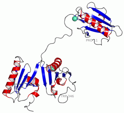
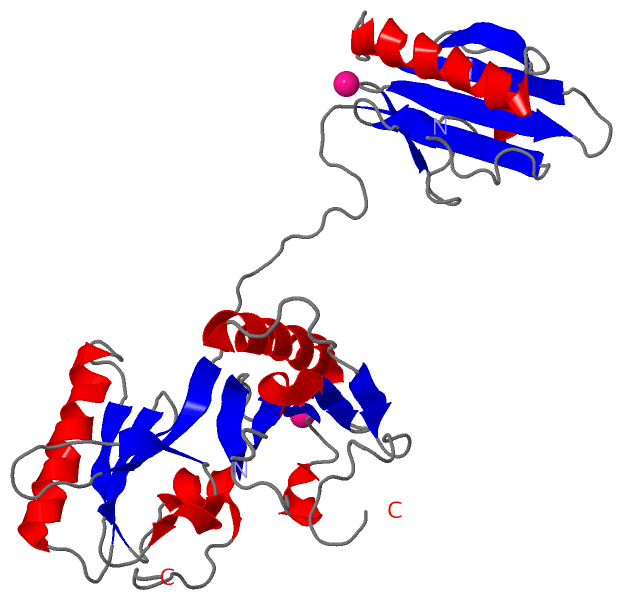
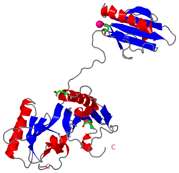
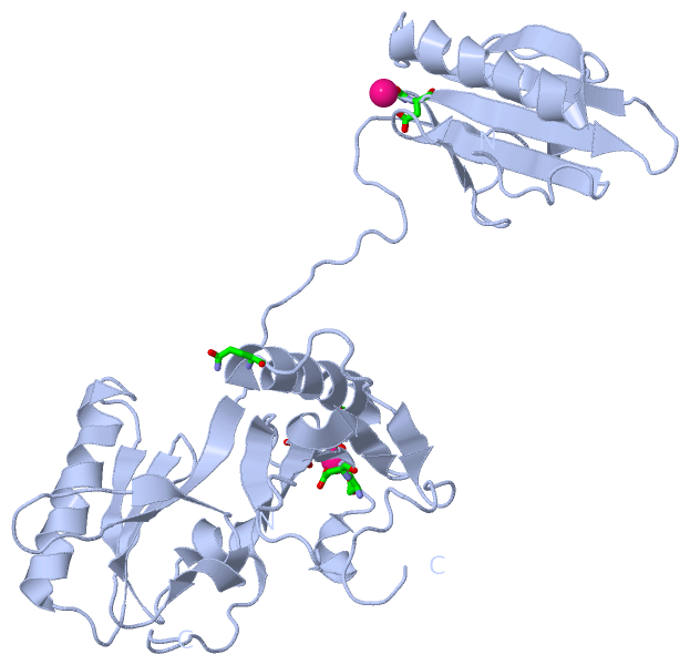
 Visualization

(-) Interactive Views

Asymmetric Unit
  Complete Structure
    Jena3D(integrated viewing of ligand, site, SAP, PROSITE, SCOP information)
    WebMol | AstexViewer[tm]@PDBe
(Java Applets, require no local installation except for Java; loading may be slow)
    STRAP
(Java WebStart application, automatic local installation, requires Java; full application with system access!)
    RasMol
(require local installation)
    Molscript (VRML)
(requires installation of a VRML viewer; select preferred view via VRML and generate a mono or stereo PDF format file)
 
  Ligands, Modified Residues, Ions
    EU3  [ RasMol | Jena3D ]  +environment [ RasMol | Jena3D ]
 
  Sites
    AC1  [ RasMol ]  +environment [ RasMol ]
    AC2  [ RasMol ]  +environment [ RasMol ]
 
  Cis Peptide Bonds
(no "Cis Peptide Bonds" information available for 1jhw)
 
Biological Units
  Complete Structure
    Biological Unit 1  [ Jena3D ]
    Biological Unit 2  [ Jena3D ]

(-) Still Images

Jmol
  protein: cartoon or spacefill or dots and stick; nucleic acid: cartoon and stick; ligands: spacefill; active site: stick
Molscript
  protein, nucleic acid: cartoon; ligands: spacefill; active site: ball and stick

 Databases and Analysis Tools

(-) Databases

Access by PDB/NDB ID
  1jhw
    Family and Domain InformationProDom | SYSTERS
    General Structural InformationGlycoscienceDB | MMDB | NDB | OCA | PDB | PDBe | PDBj | PDBsum | PDBWiki | PQS | PROTEOPEDIA
    Orientation in MembranesOPM
    Protein SurfaceSURFACE
    Secondary StructureDSSP (structure derived) | HSSP (homology derived)
    Structural GenomicsGeneCensus
    Structural NeighboursCE | VAST
    Structure ClassificationCATH | Dali | SCOP
    Validation and Original DataBMRB Data View | BMRB Restraints Grid | EDS | PROCHECK | RECOORD | WHAT_CHECK
 
Access by UniProt ID/Accession number
  CAPG_HUMAN | P40121
    Comparative Protein Structure ModelsModBase
    Genomic InformationEnsembl
    Protein-protein InteractionDIP
    Sequence, Family and Domain InformationInterPro | Pfam | SMART | UniProtKB/SwissProt
 
Access by Enzyme Classificator   (EC Number)
  (no 'Enzyme Classificator' available)
    General Enzyme InformationBRENDA | EC-PDB | Enzyme | IntEnz
    PathwayKEGG | MetaCyc
 
Access by Disease Identifier   (MIM ID)
  (no 'MIM ID' available)
    Disease InformationOMIM
 
Access by GenAge ID
  (no 'GenAge ID' available)
    Age Related InformationGenAge

(-) Analysis Tools

Access by PDB/NDB ID
    Domain InformationXDom
    Interatomic Contacts of Structural UnitsCSU
    Ligand-protein ContactsLPC
    Protein CavitiescastP
    Sequence and Secondary StructurePDBCartoon
    Structure AlignmentSTRAP(Java WebStart application, automatic local installation, requires Java; full application with system access!)
    Structure and Sequence BrowserSTING
 
Access by UniProt ID/Accession number
  CAPG_HUMAN | P40121
    Protein Disorder PredictionDisEMBL | FoldIndex | GLOBPLOT (for more information see DisProt)

 Related Entries

(-) Entries Sharing at Least One Protein Chain (UniProt ID)

UniProtKB/Swiss-Prot
        CAPG_HUMAN | P401211j72

(-) Related Entries Specified in the PDB File

1j72 1J72 IS THE CRYSTAL STRUCTURE OF MUTANT MACROPHAGE CAPPING PROTEIN (CAP G) WITH ACTIN-SEVERING ACTIVITY IN THE CA2+- FREE FORM