Show PDB file:   
         Plain Text   HTML   (compressed file size)
QuickSearch:   
by PDB,NDB,UniProt,PROSITE Code or Search Term(s)  
(-)Biol.Unit 1 - manually
(-)Asymmetric Unit
(-)Biological Unit 1
collapse expand < >
Image Biol.Unit 1 - manually
Biol.Unit 1 - manually  (Jmol Viewer)
Image Asymmetric Unit
Asymmetric Unit  (Jmol Viewer)
Image Biological Unit 1
Biological Unit 1  (Jmol Viewer)

(-) Description

Title :  CRYSTAL STRUCTURE OF 3',5"-AMINOGLYCOSIDE PHOSPHOTRANSFERASE TYPE IIIA APOENZYME
 
Authors :  D. L. Burk, W. C. Hon, A. K. -W. Leung, A. M. Berghuis
Date :  16 May 01  (Deposition) - 08 Aug 01  (Release) - 24 Feb 09  (Revision)
Method :  X-RAY DIFFRACTION
Resolution :  3.20
Chains :  Asym. Unit :  A
Biol. Unit 1:  A  (2x)
Keywords :  Antibiotic Resistance, Kinase, Atp-Binding, Transferase (Keyword Search: [Gene Ontology, PubMed, Web (Google))
 
Reference :  D. L. Burk, W. C. Hon, A. K. Leung, A. M. Berghuis
Structural Analyses Of Nucleotide Binding To An Aminoglycoside Phosphotransferase.
Biochemistry V. 40 8756 2001
PubMed-ID: 11467935  |  Reference-DOI: 10.1021/BI010504P
(for further references see the PDB file header)

(-) Compounds

Molecule 1 - AMINOGLYCOSIDE 3'-PHOSPHOTRANSFERASE
    ChainsA
    EC Number2.7.1.95
    EngineeredYES
    Expression SystemESCHERICHIA COLI BL21(DE3)
    Expression System PlasmidPPCRG6
    Expression System StrainBL21(DE3)
    Expression System Taxid469008
    Expression System Vector TypePLASMID
    Organism ScientificENTEROCOCCUS FAECALIS
    Organism Taxid1351
    SynonymAPH(3')III

 Structural Features

(-) Chains, Units

  1
Asymmetric Unit A
Biological Unit 1 (2x)A

Summary Information (see also Sequences/Alignments below)

(-) Ligands, Modified Residues, Ions  (0, 0)

(no "Ligand,Modified Residues,Ions" information available for 1J7I)

(-) Sites  (0, 0)

(no "Site" information available for 1J7I)

(-) SS Bonds  (0, 0)

(no "SS Bond" information available for 1J7I)

(-) Cis Peptide Bonds  (0, 0)

(no "Cis Peptide Bond" information available for 1J7I)

 Sequence-Structure Mapping

(-) SAPs(SNPs)/Variants  (0, 0)

(no "SAP(SNP)/Variant" information available for 1J7I)

(-) PROSITE Motifs  (0, 0)

(no "PROSITE Motif" information available for 1J7I)

(-) Exons   (0, 0)

(no "Exon" information available for 1J7I)

(-) Sequences/Alignments

Asymmetric Unit
   Reformat: Number of residues per line =  ('0' or empty: single-line sequence representation)
  Number of residues per labelling interval =   
  UniProt sequence: complete  aligned part    
   Show mapping: SCOP domains CATH domains Pfam domains Secondary structure (by author)
SAPs(SNPs) PROSITE motifs Exons
(details for a mapped element are shown in a popup box when the mouse pointer rests over it)
Chain A from PDB  Type:PROTEIN  Length:260
 aligned with KKA3_ENTFL | P0A3Y5 from UniProtKB/Swiss-Prot  Length:264

    Alignment length:260
                                    14        24        34        44        54        64        74        84        94       104       114       124       134       144       154       164       174       184       194       204       214       224       234       244       254       264
           KKA3_ENTFL     5 RISPELKKLIEKYRCVKDTEGMSPAKVYKLVGENENLYLKMTDSRYKGTTYDVEREKDMMLWLEGKLPVPKVLHFERHDGWSNLLMSEADGVLCSEEYEDEQSPEKIIELYAECIRLFHSIDISDCPYTNSLDSRLAELDYLLNNDLADVDCENWEEDTPFKDPRELYDFLKTEKPEEELVFSHGDLGDSNIFVKDGKVSGFIDLGRSGRADKWYDIAFCVRSIREDIGEEQYVELFFDLLGIKPDWEKIKYYILLDELF 264
               SCOP domains d1j7ia_ A: Type IIIa 3',5-aminoglycoside phosphotransferase                                                                                                                                                                                                          SCOP domains
               CATH domains 1j7iA01 A:5-91 Phosphorylase Kinase; domain 1                                          1j7iA02 A:92-264  [code=3.90.1200.10, no name defined]                                                                                                                        CATH domains
               Pfam domains -------------------------------------------------------------------------------------------------------------------------------------------------------------------------------------------------------------------------------------------------------------------- Pfam domains
         Sec.struct. author ..hhhhhhhhh....ee........eeeeee....eeeeeee.........hhhhhhhhhhhh........eeeeee....eeeee.....ee.........hhhhhhhhhhhhhhhhh...........hhhhhhhhhhhhhh.........hhhhhh...hhhhhhhhhhhhh....eeee........eeee..eeeee......eeeehhhhhhhhhhhhhhhh...hhhhhhhhhh....hhhhhhhhhhhhhhh Sec.struct. author
                 SAPs(SNPs) -------------------------------------------------------------------------------------------------------------------------------------------------------------------------------------------------------------------------------------------------------------------- SAPs(SNPs)
                    PROSITE -------------------------------------------------------------------------------------------------------------------------------------------------------------------------------------------------------------------------------------------------------------------- PROSITE
                 Transcript -------------------------------------------------------------------------------------------------------------------------------------------------------------------------------------------------------------------------------------------------------------------- Transcript
                 1j7i A   5 RISPELKKLIEKYRCVKDTEGMSPAKVYKLVGENENLYLKMTDSRYKGTTYDVEREKDMMLWLEGKLPVPKVLHFERHDGWSNLLMSEADGVLCSEEYEDEQSPEKIIELYAECIRLFHSIDISDCPYTNSLDSRLAELDYLLNNDLADVDCENWEEDTPFKDPRELYDFLKTEKPEEELVFSHGDLGDSNIFVKDGKVSGFIDLGRSGRADKWYDIAFCVRSIREDIGEEQYVELFFDLLGIKPDWEKIKYYILLDELF 264
                                    14        24        34        44        54        64        74        84        94       104       114       124       134       144       154       164       174       184       194       204       214       224       234       244       254       264

   Legend:   → Mismatch (orange background)
  - → Gap (green background, '-', border residues have a numbering label)
    → Modified Residue (blue background, lower-case, 'x' indicates undefined single-letter code, labelled with number + name)
  x → Chemical Group (purple background, 'x', labelled with number + name, e.g. ACE or NH2)
  extra numbering lines below/above indicate numbering irregularities and modified residue names etc., number ends below/above '|'

 Classification and Annotation

(-) SCOP Domains  (1, 1)

Asymmetric Unit

(-) CATH Domains  (2, 2)

Asymmetric Unit
(-)
Class: Alpha Beta (26913)

(-) Pfam Domains  (0, 0)

(no "Pfam Domain" information available for 1J7I)

(-) Gene Ontology  (8, 8)

Asymmetric Unit(hide GO term definitions)
Chain A   (KKA3_ENTFL | P0A3Y5)
molecular function
    GO:0005524    ATP binding    Interacting selectively and non-covalently with ATP, adenosine 5'-triphosphate, a universally important coenzyme and enzyme regulator.
    GO:0008910    kanamycin kinase activity    Catalysis of the reaction: ATP + kanamycin = ADP + 2 H(+) + kanamycin 3'-phosphate.
    GO:0016301    kinase activity    Catalysis of the transfer of a phosphate group, usually from ATP, to a substrate molecule.
    GO:0000166    nucleotide binding    Interacting selectively and non-covalently with a nucleotide, any compound consisting of a nucleoside that is esterified with (ortho)phosphate or an oligophosphate at any hydroxyl group on the ribose or deoxyribose.
    GO:0016773    phosphotransferase activity, alcohol group as acceptor    Catalysis of the transfer of a phosphorus-containing group from one compound (donor) to an alcohol group (acceptor).
    GO:0016740    transferase activity    Catalysis of the transfer of a group, e.g. a methyl group, glycosyl group, acyl group, phosphorus-containing, or other groups, from one compound (generally regarded as the donor) to another compound (generally regarded as the acceptor). Transferase is the systematic name for any enzyme of EC class 2.
biological process
    GO:0016310    phosphorylation    The process of introducing a phosphate group into a molecule, usually with the formation of a phosphoric ester, a phosphoric anhydride or a phosphoric amide.
    GO:0046677    response to antibiotic    Any process that results in a change in state or activity of a cell or an organism (in terms of movement, secretion, enzyme production, gene expression, etc.) as a result of an antibiotic stimulus. An antibiotic is a chemical substance produced by a microorganism which has the capacity to inhibit the growth of or to kill other microorganisms.
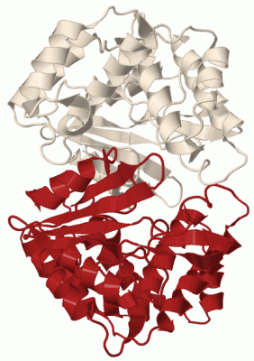
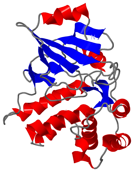
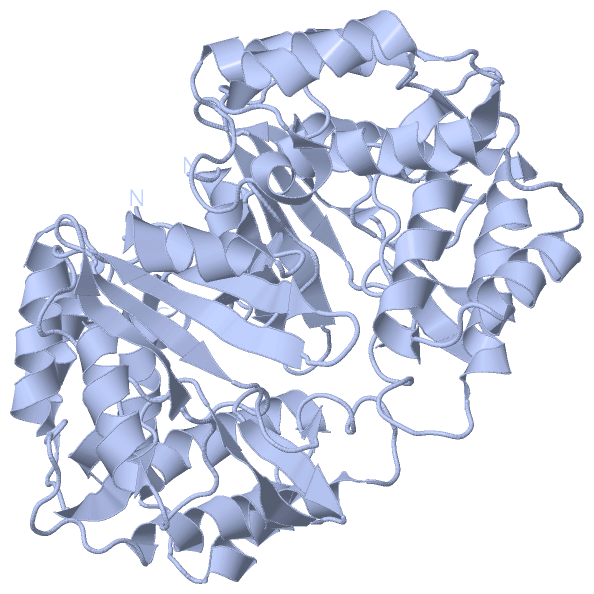
 Visualization

(-) Interactive Views

Asymmetric Unit
  Complete Structure
    Jena3D(integrated viewing of ligand, site, SAP, PROSITE, SCOP information)
    WebMol | AstexViewer[tm]@PDBe
(Java Applets, require no local installation except for Java; loading may be slow)
    STRAP
(Java WebStart application, automatic local installation, requires Java; full application with system access!)
    RasMol
(require local installation)
    Molscript (VRML)
(requires installation of a VRML viewer; select preferred view via VRML and generate a mono or stereo PDF format file)
 
  Ligands, Modified Residues, Ions
(no "Ligands, Modified Residues, Ions" information available for 1j7i)
 
  Sites
(no "Sites" information available for 1j7i)
 
  Cis Peptide Bonds
(no "Cis Peptide Bonds" information available for 1j7i)
 
Biological Unit
  Complete Structure
    Biological Unit 1  [ Jena3D ]

(-) Still Images

Jmol
  protein: cartoon or spacefill or dots and stick; nucleic acid: cartoon and stick; ligands: spacefill; active site: stick
Molscript
  protein, nucleic acid: cartoon; ligands: spacefill; active site: ball and stick

 Databases and Analysis Tools

(-) Databases

Access by PDB/NDB ID
  1j7i
    Family and Domain InformationProDom | SYSTERS
    General Structural InformationGlycoscienceDB | MMDB | NDB | OCA | PDB | PDBe | PDBj | PDBsum | PDBWiki | PQS | PROTEOPEDIA
    Orientation in MembranesOPM
    Protein SurfaceSURFACE
    Secondary StructureDSSP (structure derived) | HSSP (homology derived)
    Structural GenomicsGeneCensus
    Structural NeighboursCE | VAST
    Structure ClassificationCATH | Dali | SCOP
    Validation and Original DataBMRB Data View | BMRB Restraints Grid | EDS | PROCHECK | RECOORD | WHAT_CHECK
 
Access by UniProt ID/Accession number
  KKA3_ENTFL | P0A3Y5
    Comparative Protein Structure ModelsModBase
    Genomic InformationEnsembl
    Protein-protein InteractionDIP
    Sequence, Family and Domain InformationInterPro | Pfam | SMART | UniProtKB/SwissProt
 
Access by Enzyme Classificator   (EC Number)
  2.7.1.95
    General Enzyme InformationBRENDA | EC-PDB | Enzyme | IntEnz
    PathwayKEGG | MetaCyc
 
Access by Disease Identifier   (MIM ID)
  (no 'MIM ID' available)
    Disease InformationOMIM
 
Access by GenAge ID
  (no 'GenAge ID' available)
    Age Related InformationGenAge

(-) Analysis Tools

Access by PDB/NDB ID
    Domain InformationXDom
    Interatomic Contacts of Structural UnitsCSU
    Ligand-protein ContactsLPC
    Protein CavitiescastP
    Sequence and Secondary StructurePDBCartoon
    Structure AlignmentSTRAP(Java WebStart application, automatic local installation, requires Java; full application with system access!)
    Structure and Sequence BrowserSTING
 
Access by UniProt ID/Accession number
  KKA3_ENTFL | P0A3Y5
    Protein Disorder PredictionDisEMBL | FoldIndex | GLOBPLOT (for more information see DisProt)

 Related Entries

(-) Entries Sharing at Least One Protein Chain (UniProt ID)

UniProtKB/Swiss-Prot
        KKA3_ENTFL | P0A3Y51j7l 1j7u 1l8t 2b0q 2bkk 3q2j 3tm0

(-) Related Entries Specified in the PDB File

1j7l 1J7L CONTAINS THE THE SAME ENZYME COMPLEXED WITH ADP
1j7u 1J7U CONTAINS THE THE SAME ENZYME COMPLEXED WITH AMPPNP