|
|
|
|
 Description
|
|
Compounds
|
||||||||||||||||||||||||||||||||||||||||||||
Chains, Units
Summary Information (see also Sequences/Alignments below) |
Ligands, Modified Residues, Ions (3, 5)
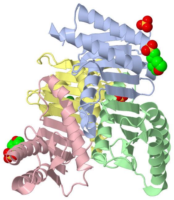
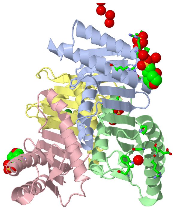
Asymmetric Unit (3, 5)
|
Sites (5, 5)
Asymmetric Unit (5, 5)
|
SS Bonds (0, 0)| (no "SS Bond" information available for 1J3W) |
Cis Peptide Bonds (0, 0)| (no "Cis Peptide Bond" information available for 1J3W) |
SAPs(SNPs)/Variants (0, 0)| (no "SAP(SNP)/Variant" information available for 1J3W) |
PROSITE Motifs (0, 0)| (no "PROSITE Motif" information available for 1J3W) |
Exons (0, 0)| (no "Exon" information available for 1J3W) |
Sequences/Alignments
Asymmetric UnitChain A from PDB Type:PROTEIN Length:134 aligned with Q9X9L0_THETH | Q9X9L0 from UniProtKB/TrEMBL Length:163 Alignment length:134 15 25 35 45 55 65 75 85 95 105 115 125 135 Q9X9L0_THETH 6 LVLYGAPYERAVEVLEETLRETGARYALLIDRKGFVLAHKEALWAPKPPPLDTLATLVAGNAAATQALAKLLGEARFQEEVHQGERMGLYVDEAGEHALLVLVFDETAPLGKVKLHGKRAAEALARIAEEALAN 139 SCOP domains d1j3wa_ A: Giding protein MglB SCOP domains CATH domains 1j3wA00 A:6-139 Dynein light chain 2a, cytoplasmic CATH domains Pfam domains -------------------------------------------------------------------------------------------------------------------------------------- Pfam domains SAPs(SNPs) -------------------------------------------------------------------------------------------------------------------------------------- SAPs(SNPs) PROSITE -------------------------------------------------------------------------------------------------------------------------------------- PROSITE Transcript -------------------------------------------------------------------------------------------------------------------------------------- Transcript 1j3w A 6 LVLYGAPYERAVEVLEETLRETGARYALLIDRKGFVLAHKEALWAPKPPPLDTLATLVAGNAAATQALAKLLGEARFQEEVHQGERMGLYVDEAGEHALLVLVFDETAPLGKVKLHGKRASEALARIAEEALAN 139 15 25 35 45 55 65 75 85 95 105 115 125 135 Chain B from PDB Type:PROTEIN Length:135 aligned with Q9X9L0_THETH | Q9X9L0 from UniProtKB/TrEMBL Length:163 Alignment length:135 11 21 31 41 51 61 71 81 91 101 111 121 131 Q9X9L0_THETH 2 VEPSLVLYGAPYERAVEVLEETLRETGARYALLIDRKGFVLAHKEALWAPKPPPLDTLATLVAGNAAATQALAKLLGEARFQEEVHQGERMGLYVDEAGEHALLVLVFDETAPLGKVKLHGKRAAEALARIAEEA 136 SCOP domains d1j3wb_ B: Giding protein MglB SCOP domains CATH domains 1j3wB00 B:2-136 Dynein light chain 2a, cytoplasmic CATH domains Pfam domains --------------------------------------------------------------------------------------------------------------------------------------- Pfam domains SAPs(SNPs) --------------------------------------------------------------------------------------------------------------------------------------- SAPs(SNPs) PROSITE --------------------------------------------------------------------------------------------------------------------------------------- PROSITE Transcript --------------------------------------------------------------------------------------------------------------------------------------- Transcript 1j3w B 2 VEPSLVLYGAPYERAVEVLEETLRETGARYALLIDRKGFVLAHKEALWAPKPPPLDTLATLVAGNAAATQALAKLLGEARFQEEVHQGERMGLYVDEAGEHALLVLVFDETAPLGKVKLHGKRASEALARIAEEA 136 11 21 31 41 51 61 71 81 91 101 111 121 131 Chain C from PDB Type:PROTEIN Length:133 aligned with Q9X9L0_THETH | Q9X9L0 from UniProtKB/TrEMBL Length:163 Alignment length:133 15 25 35 45 55 65 75 85 95 105 115 125 135 Q9X9L0_THETH 6 LVLYGAPYERAVEVLEETLRETGARYALLIDRKGFVLAHKEALWAPKPPPLDTLATLVAGNAAATQALAKLLGEARFQEEVHQGERMGLYVDEAGEHALLVLVFDETAPLGKVKLHGKRAAEALARIAEEALA 138 SCOP domains d1j3wc_ C: Giding protein MglB SCOP domains CATH domains 1j3wC00 C:6-138 Dynein light chain 2a, cytoplasmic CATH domains Pfam domains ------------------------------------------------------------------------------------------------------------------------------------- Pfam domains SAPs(SNPs) ------------------------------------------------------------------------------------------------------------------------------------- SAPs(SNPs) PROSITE ------------------------------------------------------------------------------------------------------------------------------------- PROSITE Transcript ------------------------------------------------------------------------------------------------------------------------------------- Transcript 1j3w C 6 LVLYGAPYERAVEVLEETLRETGARYALLIDRKGFVLAHKEALWAPKPPPLDTLATLVAGNAAATQALAKLLGEARFQEEVHQGERMGLYVDEAGEHALLVLVFDETAPLGKVKLHGKRASEALARIAEEALA 138 15 25 35 45 55 65 75 85 95 105 115 125 135 Chain D from PDB Type:PROTEIN Length:133 aligned with Q9X9L0_THETH | Q9X9L0 from UniProtKB/TrEMBL Length:163 Alignment length:133 15 25 35 45 55 65 75 85 95 105 115 125 135 Q9X9L0_THETH 6 LVLYGAPYERAVEVLEETLRETGARYALLIDRKGFVLAHKEALWAPKPPPLDTLATLVAGNAAATQALAKLLGEARFQEEVHQGERMGLYVDEAGEHALLVLVFDETAPLGKVKLHGKRAAEALARIAEEALA 138 SCOP domains d1j3wd_ D: Giding protein MglB SCOP domains CATH domains 1j3wD00 D:6-138 Dynein light chain 2a, cytoplasmic CATH domains Pfam domains ------------------------------------------------------------------------------------------------------------------------------------- Pfam domains SAPs(SNPs) ------------------------------------------------------------------------------------------------------------------------------------- SAPs(SNPs) PROSITE ------------------------------------------------------------------------------------------------------------------------------------- PROSITE Transcript ------------------------------------------------------------------------------------------------------------------------------------- Transcript 1j3w D 6 LVLYGAPYERAVEVLEETLRETGARYALLIDRKGFVLAHKEALWAPKPPPLDTLATLVAGNAAATQALAKLLGEARFQEEVHQGERMGLYVDEAGEHALLVLVFDETAPLGKVKLHGKRASEALARIAEEALA 138 15 25 35 45 55 65 75 85 95 105 115 125 135
|
||||||||||||||||||||
SCOP Domains (1, 4)
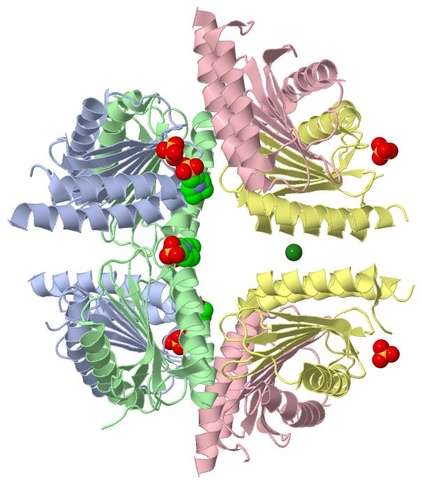
Asymmetric Unit
 |
CATH Domains (1, 4)
Asymmetric Unit
|
Pfam Domains (0, 0)| (no "Pfam Domain" information available for 1J3W) |
Gene Ontology (0, 0)|
Asymmetric Unit(hide GO term definitions)
(no "Gene Ontology" information available for 1J3W)
|
Interactive Views
|
|||||||||||||||||||||||||||||||||||||||||||||||||||||||||||||||||||||||||||||||||||||||||||||||||||||||||||||||||||||||||||||||||||||||||||||||||||||||||||||||||||||||||||||||||||||||
Still Images
|
||||||||||||||||
Databases
|
||||||||||||||||||||||||||||||||||||||||||||||||||||||||||||||||||||||||||||||||||||||||||||||||||||||||||||||||||||||||||||||||||||||||||||||||||||||||||||||||
Analysis Tools
|
|||||||||||||||||||||||||||||||||||||||||||||||||||||||||||||
Entries Sharing at Least One Protein Chain (UniProt ID)
Related Entries Specified in the PDB File
|
|