| molecular function |
|---|
| | GO:0005524 | | ATP binding | | Interacting selectively and non-covalently with ATP, adenosine 5'-triphosphate, a universally important coenzyme and enzyme regulator. |
| | GO:0016301 | | kinase activity | | Catalysis of the transfer of a phosphate group, usually from ATP, to a substrate molecule. |
| | GO:0000166 | | nucleotide binding | | Interacting selectively and non-covalently with a nucleotide, any compound consisting of a nucleoside that is esterified with (ortho)phosphate or an oligophosphate at any hydroxyl group on the ribose or deoxyribose. |
| | GO:0005515 | | protein binding | | Interacting selectively and non-covalently with any protein or protein complex (a complex of two or more proteins that may include other nonprotein molecules). |
| | GO:0019904 | | protein domain specific binding | | Interacting selectively and non-covalently with a specific domain of a protein. |
| | GO:0004672 | | protein kinase activity | | Catalysis of the phosphorylation of an amino acid residue in a protein, usually according to the reaction: a protein + ATP = a phosphoprotein + ADP. |
| | GO:0004674 | | protein serine/threonine kinase activity | | Catalysis of the reactions: ATP + protein serine = ADP + protein serine phosphate, and ATP + protein threonine = ADP + protein threonine phosphate. |
| | GO:0016740 | | transferase activity | | Catalysis of the transfer of a group, e.g. a methyl group, glycosyl group, acyl group, phosphorus-containing, or other groups, from one compound (generally regarded as the donor) to another compound (generally regarded as the acceptor). Transferase is the systematic name for any enzyme of EC class 2. |
| biological process |
|---|
| | GO:0008063 | | Toll signaling pathway | | A series of molecular signals initiated by the binding of an extracellular ligand to the receptor Toll on the surface of a target cell, and ending with regulation of a downstream cellular process, e.g. transcription. |
| | GO:0019732 | | antifungal humoral response | | An immune response against a fungus mediated through a body fluid. An example of this process is the antifungal humoral response in Drosophila melanogaster. |
| | GO:0006915 | | apoptotic process | | A programmed cell death process which begins when a cell receives an internal (e.g. DNA damage) or external signal (e.g. an extracellular death ligand), and proceeds through a series of biochemical events (signaling pathway phase) which trigger an execution phase. The execution phase is the last step of an apoptotic process, and is typically characterized by rounding-up of the cell, retraction of pseudopodes, reduction of cellular volume (pyknosis), chromatin condensation, nuclear fragmentation (karyorrhexis), plasma membrane blebbing and fragmentation of the cell into apoptotic bodies. When the execution phase is completed, the cell has died. |
| | GO:0006952 | | defense response | | Reactions, triggered in response to the presence of a foreign body or the occurrence of an injury, which result in restriction of damage to the organism attacked or prevention/recovery from the infection caused by the attack. |
| | GO:0048262 | | determination of dorsal/ventral asymmetry | | Determination of asymmetry from the dorsal to the ventral side; as, the dorsoventral axis. |
| | GO:0009950 | | dorsal/ventral axis specification | | The establishment, maintenance and elaboration of the dorsal/ventral axis. The dorsal/ventral axis is defined by a line that runs orthogonal to both the anterior/posterior and left/right axes. The dorsal end is defined by the upper or back side of an organism. The ventral end is defined by the lower or front side of an organism. |
| | GO:0035172 | | hemocyte proliferation | | The multiplication or reproduction of hemocytes, resulting in the expansion of the cell population. Hemocytes are blood cells associated with a hemocoel (the cavity containing most of the major organs of the arthropod body) which are involved in defense and clotting of hemolymph, but not involved in transport of oxygen. |
| | GO:0030097 | | hemopoiesis | | The process whose specific outcome is the progression of the myeloid and lymphoid derived organ/tissue systems of the blood and other parts of the body over time, from formation to the mature structure. The site of hemopoiesis is variable during development, but occurs primarily in bone marrow or kidney in many adult vertebrates. |
| | GO:0045087 | | innate immune response | | Innate immune responses are defense responses mediated by germline encoded components that directly recognize components of potential pathogens. |
| | GO:0035556 | | intracellular signal transduction | | The process in which a signal is passed on to downstream components within the cell, which become activated themselves to further propagate the signal and finally trigger a change in the function or state of the cell. |
| | GO:0016310 | | phosphorylation | | The process of introducing a phosphate group into a molecule, usually with the formation of a phosphoric ester, a phosphoric anhydride or a phosphoric amide. |
| | GO:0046777 | | protein autophosphorylation | | The phosphorylation by a protein of one or more of its own amino acid residues (cis-autophosphorylation), or residues on an identical protein (trans-autophosphorylation). |
| | GO:0006606 | | protein import into nucleus | | The directed movement of a protein from the cytoplasm to the nucleus. |
| | GO:0006468 | | protein phosphorylation | | The process of introducing a phosphate group on to a protein. |
| | GO:0030162 | | regulation of proteolysis | | Any process that modulates the frequency, rate or extent of the hydrolysis of a peptide bond or bonds within a protein. |
| | GO:0009620 | | response to fungus | | Any process that results in a change in state or activity of a cell or an organism (in terms of movement, secretion, enzyme production, gene expression, etc.) as a result of a stimulus from a fungus. |
| | GO:0007165 | | signal transduction | | The cellular process in which a signal is conveyed to trigger a change in the activity or state of a cell. Signal transduction begins with reception of a signal (e.g. a ligand binding to a receptor or receptor activation by a stimulus such as light), or for signal transduction in the absence of ligand, signal-withdrawal or the activity of a constitutively active receptor. Signal transduction ends with regulation of a downstream cellular process, e.g. regulation of transcription or regulation of a metabolic process. Signal transduction covers signaling from receptors located on the surface of the cell and signaling via molecules located within the cell. For signaling between cells, signal transduction is restricted to events at and within the receiving cell. |
| | GO:0007352 | | zygotic specification of dorsal/ventral axis | | The specification of the dorsal/ventral axis of the embryo, through the products of genes expressed in the zygote. |
| cellular component |
|---|
| | GO:0005737 | | cytoplasm | | All of the contents of a cell excluding the plasma membrane and nucleus, but including other subcellular structures. |
| | GO:0016020 | | membrane | | A lipid bilayer along with all the proteins and protein complexes embedded in it an attached to it. |
| | GO:0005634 | | nucleus | | A membrane-bounded organelle of eukaryotic cells in which chromosomes are housed and replicated. In most cells, the nucleus contains all of the cell's chromosomes except the organellar chromosomes, and is the site of RNA synthesis and processing. In some species, or in specialized cell types, RNA metabolism or DNA replication may be absent. |
| | GO:0005886 | | plasma membrane | | The membrane surrounding a cell that separates the cell from its external environment. It consists of a phospholipid bilayer and associated proteins. |
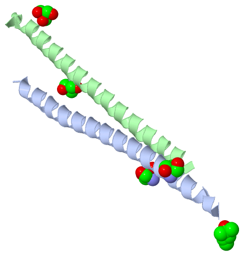
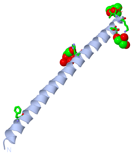
 Description
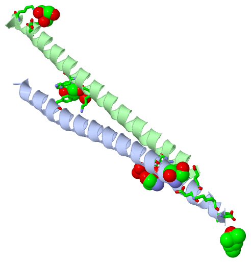
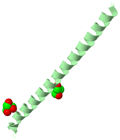