|
|
|
|
 Description
|
|
Compounds
|
||||||||||||||||||||||||||||||||||||||||||||||||||||||||||||||||||||
Chains, Units
Summary Information (see also Sequences/Alignments below) |
Ligands, Modified Residues, Ions (1, 1)
NMR Structure (1, 1)
|
Sites (1, 1)
NMR Structure (1, 1)
|
SS Bonds (0, 0)| (no "SS Bond" information available for 1I6D) |
Cis Peptide Bonds (0, 0)| (no "Cis Peptide Bond" information available for 1I6D) |
SAPs(SNPs)/Variants (0, 0)| (no "SAP(SNP)/Variant" information available for 1I6D) |
PROSITE Motifs (1, 1)
NMR Structure (1, 1)
|
||||||||||||||||||||||||
Exons (0, 0)| (no "Exon" information available for 1I6D) |
Sequences/Alignments
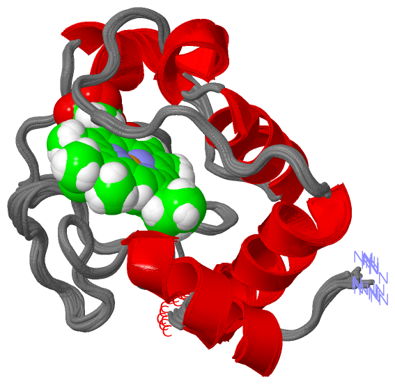
NMR StructureChain A from PDB Type:PROTEIN Length:100 aligned with CY552_PARDE | P54820 from UniProtKB/Swiss-Prot Length:176 Alignment length:100 86 96 106 116 126 136 146 156 166 176 CY552_PARDE 77 SADPAAGEKVFGKCKACHKLDGNDGVGPHLNGVVGRTVAGVDGFNYSDPMKAHGGDWTPEALQEFLTNPKAVVKGTKMAFAGLPKIEDRANLIAYLEGQQ 176 SCOP domains d1i6da_ A: Cytochrome c552 SCOP domains CATH domains 1i6dA00 A:1-100 Cytochrome c CATH domains Pfam domains ---------------------------------------------------------------------------------------------------- Pfam domains SAPs(SNPs) ---------------------------------------------------------------------------------------------------- SAPs(SNPs) PROSITE -CYTC PDB: A:2-100 UniProt: 78-176 PROSITE Transcript ---------------------------------------------------------------------------------------------------- Transcript 1i6d A 1 MADPAAGEKVFGKCKACHKLDGNDGVGPHLNGVVGRTVAGVDGFNYSDPMKAHGGDWTPEALQEFLTNPKAVVKGTKMAFAGLPKIEDRANLIAYLEGQQ 100 10 20 30 40 50 60 70 80 90 100
|
||||||||||||||||||||
SCOP Domains (1, 1)
NMR Structure
 |
CATH Domains (1, 1)
NMR Structure
|
Pfam Domains (0, 0)| (no "Pfam Domain" information available for 1I6D) |
Gene Ontology (7, 7)|
NMR Structure(hide GO term definitions) Chain A (CY552_PARDE | P54820)
|
||||||||||||||||||||||||||||||||||||||||||||||||||||||||||||
Interactive Views
|
||||||||||||||||||||||||||||||||||||||||||||||||||||||||||||||||||||||||||||||||||||||||||||||||||||||||||||||||||||||
Still Images
|
||||||||||||||||
Databases
|
||||||||||||||||||||||||||||||||||||||||||||||||||||||||||||||||||||||||||||||||||||||||||||||||||||||||||||||||||||||||||||||||||||||||||||||||||||||||||||||||
Analysis Tools
|
|||||||||||||||||||||||||||||||||||||||||||||||||||||||||||||
Entries Sharing at Least One Protein Chain (UniProt ID)
Related Entries Specified in the PDB File
|
|