|
|
|
|
 Description
|
|
Compounds
|
||||||||||||||||||||||||||||||||||||||||||||||||
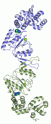
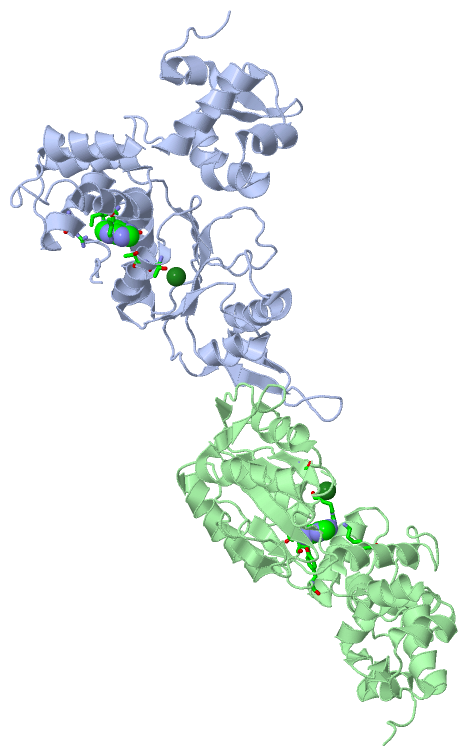
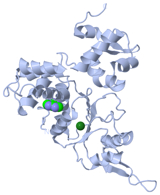
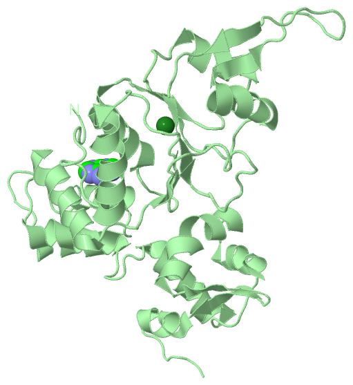
Chains, Units
Summary Information (see also Sequences/Alignments below) |
Ligands, Modified Residues, Ions (2, 4)| Asymmetric Unit (2, 4) Biological Unit 1 (1, 1) Biological Unit 2 (1, 1) |
Sites (4, 4)
Asymmetric Unit (4, 4)
|
SS Bonds (0, 0)| (no "SS Bond" information available for 1HQC) |
Cis Peptide Bonds (0, 0)| (no "Cis Peptide Bond" information available for 1HQC) |
SAPs(SNPs)/Variants (0, 0)| (no "SAP(SNP)/Variant" information available for 1HQC) |
PROSITE Motifs (0, 0)| (no "PROSITE Motif" information available for 1HQC) |
Exons (0, 0)| (no "Exon" information available for 1HQC) |
Sequences/Alignments
Asymmetric UnitChain A from PDB Type:PROTEIN Length:314 aligned with RUVB_THET8 | Q5SL87 from UniProtKB/Swiss-Prot Length:324 Alignment length:314 14 24 34 44 54 64 74 84 94 104 114 124 134 144 154 164 174 184 194 204 214 224 234 244 254 264 274 284 294 304 314 RUVB_THET8 5 ALRPKTLDEYIGQERLKQKLRVYLEAAKARKEPLEHLLLFGPPGLGKTTLAHVIAHELGVNLRVTSGPAIEKPGDLAAILANSLEEGDILFIDEIHRLSRQAEEHLYPAMEDFVMDIVIGQGPAARTIRLELPRFTLIGATTRPGLITAPLLSRFGIVEHLEYYTPEELAQGVMRDARLLGVRITEEAALEIGRRSRGTMRVAKRLFRRVRDFAQVAGEEVITRERALEALAALGLDELGLEKRDREILEVLILRFGGGPVGLATLATALSEDPGTLEEVHEPYLIRQGLLKRTPRGRVATELAYRHLGYPPPV 318 SCOP domains d1hqca2 A:5-242 Holliday junction helicase RuvB d1hqca1 A:243-318 Holliday junction helicase RuvB SCOP domains CATH domains 1hqcA01 A:5-167 P-loop containing nucleotide triphosphate hydrolases 1hqcA02 A:168-243 [code=1.10.8.60, no name defined] 1hqcA03 A:244-318 'winged helix' repressor DNA binding domain CATH domains Pfam domains -------------------------------------------------------------------------------------------------------------------------------------------------------------------------------------------------------------------------------------------------------------------------------------------------------------------------- Pfam domains SAPs(SNPs) -------------------------------------------------------------------------------------------------------------------------------------------------------------------------------------------------------------------------------------------------------------------------------------------------------------------------- SAPs(SNPs) PROSITE -------------------------------------------------------------------------------------------------------------------------------------------------------------------------------------------------------------------------------------------------------------------------------------------------------------------------- PROSITE Transcript -------------------------------------------------------------------------------------------------------------------------------------------------------------------------------------------------------------------------------------------------------------------------------------------------------------------------- Transcript 1hqc A 5 ALRPKTLDEYIGQERLKQKLRVYLEAAKARKEPLEHLLLFGPPGLGKTTLAHVIAHELGVNLRVTSGPAIEKPGDLAAILANSLEEGDILFIDEIHRLSRQAEEHLYPAMEDFVMDIVIGQGPAARTIRLELPRFTLIGATTRPGLITAPLLSRFGIVEHLEYYTPEELAQGVMRDARLLGVRITEEAALEIGRRSRGTMRVAKRLFRRVRDFAQVAGEEVITRERALEALAALGLDELGLEKRDREILEVLILRFGGGPVGLATLATALSEDPGTLEEVHEPYLIRQGLLKRTPRGRVPTELAYRHLGYPPPV 318 14 24 34 44 54 64 74 84 94 104 114 124 134 144 154 164 174 184 194 204 214 224 234 244 254 264 274 284 294 304 314 Chain B from PDB Type:PROTEIN Length:314 aligned with RUVB_THET8 | Q5SL87 from UniProtKB/Swiss-Prot Length:324 Alignment length:314 14 24 34 44 54 64 74 84 94 104 114 124 134 144 154 164 174 184 194 204 214 224 234 244 254 264 274 284 294 304 314 RUVB_THET8 5 ALRPKTLDEYIGQERLKQKLRVYLEAAKARKEPLEHLLLFGPPGLGKTTLAHVIAHELGVNLRVTSGPAIEKPGDLAAILANSLEEGDILFIDEIHRLSRQAEEHLYPAMEDFVMDIVIGQGPAARTIRLELPRFTLIGATTRPGLITAPLLSRFGIVEHLEYYTPEELAQGVMRDARLLGVRITEEAALEIGRRSRGTMRVAKRLFRRVRDFAQVAGEEVITRERALEALAALGLDELGLEKRDREILEVLILRFGGGPVGLATLATALSEDPGTLEEVHEPYLIRQGLLKRTPRGRVATELAYRHLGYPPPV 318 SCOP domains d1hqcb2 B:5-242 Holliday junction helicase RuvB d1hqcb1 B:243-318 Holliday junction helicase RuvB SCOP domains CATH domains 1hqcB01 B:5-167 P-loop containing nucleotide triphosphate hydrolases 1hqcB02 B:168-243 [code=1.10.8.60, no name defined] 1hqcB03 B:244-318 'winged helix' repressor DNA binding domain CATH domains Pfam domains -------------------------------------------------------------------------------------------------------------------------------------------------------------------------------------------------------------------------------------------------------------------------------------------------------------------------- Pfam domains SAPs(SNPs) -------------------------------------------------------------------------------------------------------------------------------------------------------------------------------------------------------------------------------------------------------------------------------------------------------------------------- SAPs(SNPs) PROSITE -------------------------------------------------------------------------------------------------------------------------------------------------------------------------------------------------------------------------------------------------------------------------------------------------------------------------- PROSITE Transcript -------------------------------------------------------------------------------------------------------------------------------------------------------------------------------------------------------------------------------------------------------------------------------------------------------------------------- Transcript 1hqc B 5 ALRPKTLDEYIGQERLKQKLRVYLEAAKARKEPLEHLLLFGPPGLGKTTLAHVIAHELGVNLRVTSGPAIEKPGDLAAILANSLEEGDILFIDEIHRLSRQAEEHLYPAMEDFVMDIVIGQGPAARTIRLELPRFTLIGATTRPGLITAPLLSRFGIVEHLEYYTPEELAQGVMRDARLLGVRITEEAALEIGRRSRGTMRVAKRLFRRVRDFAQVAGEEVITRERALEALAALGLDELGLEKRDREILEVLILRFGGGPVGLATLATALSEDPGTLEEVHEPYLIRQGLLKRTPRGRVPTELAYRHLGYPPPV 318 14 24 34 44 54 64 74 84 94 104 114 124 134 144 154 164 174 184 194 204 214 224 234 244 254 264 274 284 294 304 314
|
||||||||||||||||||||
SCOP Domains (2, 4)
Asymmetric Unit
 |
CATH Domains (3, 6)
Asymmetric Unit
|
Pfam Domains (0, 0)| (no "Pfam Domain" information available for 1HQC) |
Gene Ontology (11, 11)|
Asymmetric Unit(hide GO term definitions) Chain A,B (RUVB_THET8 | Q5SL87)
|
||||||||||||||||||||||||||||||||||||||||||||||||||||||||||||||||||||||||||||||
Interactive Views
|
|||||||||||||||||||||||||||||||||||||||||||||||||||||||||||||||||||||||||||||||||||||||||||||||||||||||||||||||||||||||||||||||||||||||||||||||||||||||||||||||||||||||||
Still Images
|
||||||||||||||||
Databases
|
||||||||||||||||||||||||||||||||||||||||||||||||||||||||||||||||||||||||||||||||||||||||||||||||||||||||||||||||||||||||||||||||||||||||||||||||||||||||||||||||
Analysis Tools
|
|||||||||||||||||||||||||||||||||||||||||||||||||||||||||||||
Entries Sharing at Least One Protein Chain (UniProt ID)
Related Entries Specified in the PDB File
|
|