|
|
|
|
 Description
|
|
Compounds
|
||||||||||||||||
Chains, Units
Summary Information (see also Sequences/Alignments below) |
Ligands, Modified Residues, Ions (1, 1)
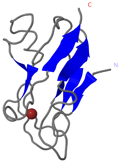
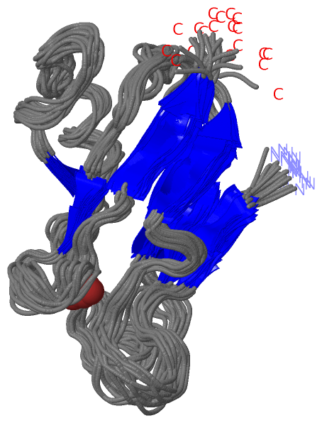
NMR Structure (1, 1)
|
Sites (1, 1)
NMR Structure (1, 1)
|
SS Bonds (0, 0)| (no "SS Bond" information available for 1FA4) |
Cis Peptide Bonds (2, 40)
NMR Structure
|
|||||||||||||||
SAPs(SNPs)/Variants (0, 0)| (no "SAP(SNP)/Variant" information available for 1FA4) |
PROSITE Motifs (1, 1)
NMR Structure (1, 1)
|
||||||||||||||||||||||||||||
Exons (0, 0)| (no "Exon" information available for 1FA4) |
Sequences/Alignments
NMR StructureChain A from PDB Type:PROTEIN Length:105 aligned with PLAS_ANAVA | P0C178 from UniProtKB/Swiss-Prot Length:105 Alignment length:105 10 20 30 40 50 60 70 80 90 100 PLAS_ANAVA 1 ETYTVKLGSDKGLLVFEPAKLTIKPGDTVEFLNNKVPPHNVVFDAALNPAKSADLAKSLSHKQLLMSPGQSTSTTFPADAPAGEYTFYCEPHRGAGMVGKITVAG 105 SCOP domains d1fa4a_ A: Plastocyanin SCOP domains CATH domains 1fa4A00 A:1-105 Cupredoxins - blue copper proteins CATH domains Pfam domains --------------------------------------------------------------------------------------------------------- Pfam domains SAPs(SNPs) --------------------------------------------------------------------------------------------------------- SAPs(SNPs) PROSITE ---------------------------------------------------------------------------------COPPER_BLUE -------- PROSITE Transcript --------------------------------------------------------------------------------------------------------- Transcript 1fa4 A 1 ETYTVKLGSDKGLLVFEPAKLTIKPGDTVEFLNNKVPPHNVVFDAALNPAKSADLAKSLSHKQLLMSPGQSTSTTFPADAPAGEYTFYCEPHRGAGMVGKITVAG 105 10 20 30 40 50 60 70 80 90 100 Chain A from PDB Type:PROTEIN Length:105 aligned with PLAS_ANAVT | Q3M9H8 from UniProtKB/Swiss-Prot Length:139 Alignment length:105 44 54 64 74 84 94 104 114 124 134 PLAS_ANAVT 35 ETYTVKLGSDKGLLVFEPAKLTIKPGDTVEFLNNKVPPHNVVFDATLNPAKSADLAKSLSHKQLLMSPGQSTSTTFPADAPAGDYSFYCEPHRGAGMVGKITVAS 139 SCOP domains d1fa4a_ A: Plastocyanin SCOP domains CATH domains 1fa4A00 A:1-105 Cupredoxins - blue copper proteins CATH domains Pfam domains --------------------------------------------------------------------------------------------------------- Pfam domains SAPs(SNPs) --------------------------------------------------------------------------------------------------------- SAPs(SNPs) PROSITE ---------------------------------------------------------------------------------COPPER_BLUE -------- PROSITE Transcript --------------------------------------------------------------------------------------------------------- Transcript 1fa4 A 1 ETYTVKLGSDKGLLVFEPAKLTIKPGDTVEFLNNKVPPHNVVFDAALNPAKSADLAKSLSHKQLLMSPGQSTSTTFPADAPAGEYTFYCEPHRGAGMVGKITVAG 105 10 20 30 40 50 60 70 80 90 100
|
||||||||||||||||||||
SCOP Domains (1, 1)
NMR Structure
 |
CATH Domains (1, 1)
NMR Structure
|
Pfam Domains (0, 0)| (no "Pfam Domain" information available for 1FA4) |
Gene Ontology (7, 14)|
NMR Structure(hide GO term definitions) Chain A (PLAS_ANAVA | P0C178)
Chain A (PLAS_ANAVT | Q3M9H8)
|
||||||||||||||||||||||||||||||||||||||||||||||||||||||||||||||||||||||||||||||||||||||||||||||||||||||||||||||||||||||||
Interactive Views
|
||||||||||||||||||||||||||||||||||||||||||||||||||||||||||||||||||||||||||||||||||||||||||||||||||||||||||||||||||||||||||||||
Still Images
|
||||||||||||||||
Databases
|
||||||||||||||||||||||||||||||||||||||||||||||||||||||||||||||||||||||||||||||||||||||||||||||||||||||||||||||||||||||||||||||||||||||||||||||||||||||||||||||||||||||||||||||||||||||||||
Analysis Tools
|
||||||||||||||||||||||||||||||||||||||||||||||||||||||||||||||||||||||||
Entries Sharing at Least One Protein Chain (UniProt ID)
Related Entries Specified in the PDB File
|
|