|
|
|
|
 Description
|
|
Compounds
|
||||||||||||||||||||||||||||
Chains, Units
Summary Information (see also Sequences/Alignments below) |
Ligands, Modified Residues, Ions (0, 0)| (no "Ligand,Modified Residues,Ions" information available for 1F2K) |
Sites (0, 0)| (no "Site" information available for 1F2K) |
SS Bonds (0, 0)| (no "SS Bond" information available for 1F2K) |
Cis Peptide Bonds (0, 0)| (no "Cis Peptide Bond" information available for 1F2K) |
SAPs(SNPs)/Variants (0, 0)| (no "SAP(SNP)/Variant" information available for 1F2K) |
PROSITE Motifs (0, 0)| (no "PROSITE Motif" information available for 1F2K) |
Exons (0, 0)| (no "Exon" information available for 1F2K) |
Sequences/Alignments
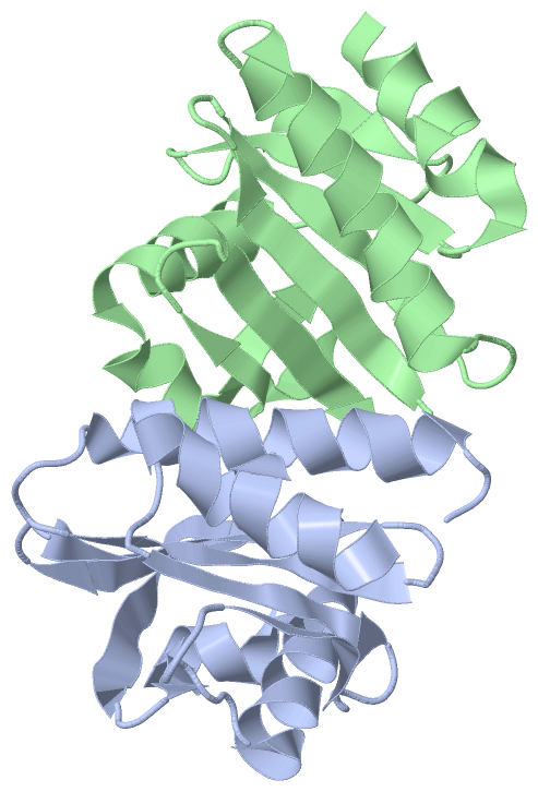
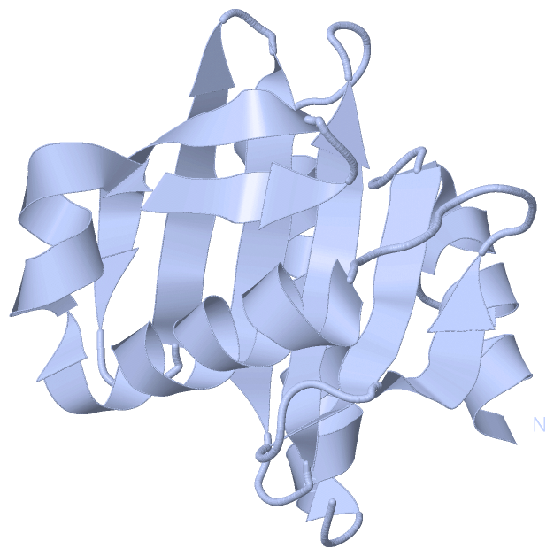
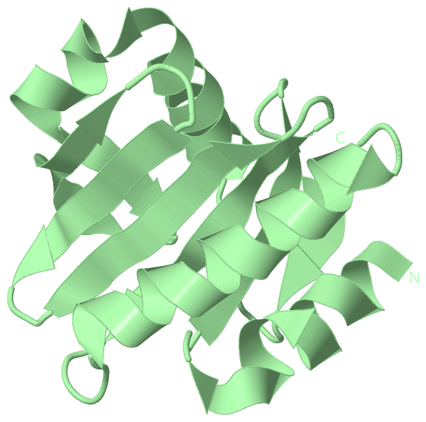
Asymmetric UnitChain A from PDB Type:PROTEIN Length:125 aligned with PROF2_ACACA | P19984 from UniProtKB/Swiss-Prot Length:126 Alignment length:125 11 21 31 41 51 61 71 81 91 101 111 121 PROF2_ACACA 2 SWQTYVDTNLVGTGAVTQAAIIGHDGNTWATSAGFAVSPANGAALANAFKDATAIRSNGFELAGTRYVTIRADDRSVYGKKGSAGVITVKTSKAILIGVYNEKIQPGTAANVVEKLADYLIGQGF 126 SCOP domains d1f2ka_ A: Profilin (actin-binding protein) SCOP domains CATH domains 1f2kA00 A:1001-1125 Dynein light chain 2a, cytoplasmic CATH domains Pfam domains ----------------------------------------------------------------------------------------------------------------------------- Pfam domains SAPs(SNPs) ----------------------------------------------------------------------------------------------------------------------------- SAPs(SNPs) PROSITE ----------------------------------------------------------------------------------------------------------------------------- PROSITE Transcript ----------------------------------------------------------------------------------------------------------------------------- Transcript 1f2k A 1001 SWQTYVDTNLVGTGAVTQAAIIGHDGNTWATSAGFAVSPANGAALANAFKDATAIRSNGFELAGTRYVTIRADDRSVYGKKGSAGVITVKTSKAILIGVYNEKIQPGTAANVVEKLADYLIGQGF 1125 1010 1020 1030 1040 1050 1060 1070 1080 1090 1100 1110 1120 Chain B from PDB Type:PROTEIN Length:125 aligned with PROF2_ACACA | P19984 from UniProtKB/Swiss-Prot Length:126 Alignment length:125 11 21 31 41 51 61 71 81 91 101 111 121 PROF2_ACACA 2 SWQTYVDTNLVGTGAVTQAAIIGHDGNTWATSAGFAVSPANGAALANAFKDATAIRSNGFELAGTRYVTIRADDRSVYGKKGSAGVITVKTSKAILIGVYNEKIQPGTAANVVEKLADYLIGQGF 126 SCOP domains d1f2kb_ B: Profilin (actin-binding protein) SCOP domains CATH domains 1f2kB00 B:2001-2125 Dynein light chain 2a, cytoplasmic CATH domains Pfam domains ----------------------------------------------------------------------------------------------------------------------------- Pfam domains SAPs(SNPs) ----------------------------------------------------------------------------------------------------------------------------- SAPs(SNPs) PROSITE ----------------------------------------------------------------------------------------------------------------------------- PROSITE Transcript ----------------------------------------------------------------------------------------------------------------------------- Transcript 1f2k B 2001 SWQTYVDTNLVGTGAVTQAAIIGHDGNTWATSAGFAVSPANGAALANAFKDATAIRSNGFELAGTRYVTIRADDRSVYGKKGSAGVITVKTSKAILIGVYNEKIQPGTAANVVEKLADYLIGQGF 2125 2010 2020 2030 2040 2050 2060 2070 2080 2090 2100 2110 2120
|
||||||||||||||||||||
SCOP Domains (1, 2)
Asymmetric Unit
 |
CATH Domains (1, 2)
Asymmetric Unit
|
Pfam Domains (0, 0)| (no "Pfam Domain" information available for 1F2K) |
Gene Ontology (3, 3)|
Asymmetric Unit(hide GO term definitions) Chain A,B (PROF2_ACACA | P19984)
|
||||||||||||||||||||||||||||||
Interactive Views
|
|||||||||||||||||||||||||||||||||||||||||||||||||||||||||||||||||||||||||||||||||||||||||||||||||||||||||||||||||||||||||||||||||||||||||||
Still Images
|
||||||||||||||||
Databases
|
||||||||||||||||||||||||||||||||||||||||||||||||||||||||||||||||||||||||||||||||||||||||||||||||||||||||||||||||||||||||||||||||||||||||||||||||||||||||||||||||
Analysis Tools
|
|||||||||||||||||||||||||||||||||||||||||||||||||||||||||||||
Entries Sharing at Least One Protein Chain (UniProt ID)
Related Entries Specified in the PDB File
|
|