Show PDB file:   
         Plain Text   HTML   (compressed file size)
QuickSearch:   
by PDB,NDB,UniProt,PROSITE Code or Search Term(s)  
(-)Asymmetric Unit
(-)Biological Unit 1
collapse expand < >
Image Asymmetric Unit
Asymmetric Unit  (Jmol Viewer)
Image Biological Unit 1
Biological Unit 1  (Jmol Viewer)

(-) Description

Title :  CRYSTAL STRUCTURE OF HUMAN MONOCYTE CHEMOTACTIC PROTEIN-2
 
Authors :  J. Blaszczyk, X. Ji
Date :  10 Apr 00  (Deposition) - 06 Dec 00  (Release) - 24 Feb 09  (Revision)
Method :  X-RAY DIFFRACTION
Resolution :  2.00
Chains :  Asym. Unit :  A
Biol. Unit 1:  A  (2x)
Keywords :  Cytokine, Chemokine, Monocyte Chemoattractant Protein, Hiv- 1, Pyroglutamic Acid, X-Ray Crystallography (Keyword Search: [Gene Ontology, PubMed, Web (Google))
 
Reference :  J. Blaszczyk, E. V. Coillie, P. Proost, J. V. Damme, G. Opdenakker, G. D. Bujacz, J. M. Wang, X. Ji
Complete Crystal Structure Of Monocyte Chemotactic Protein-2, A Cc Chemokine That Interacts With Multiple Receptors.
Biochemistry V. 39 14075 2000
PubMed-ID: 11087354  |  Reference-DOI: 10.1021/BI0009340
(for further references see the PDB file header)

(-) Compounds

Molecule 1 - MONOCYTE CHEMOTACTIC PROTEIN 2
    ChainsA
    EngineeredYES
    Expression SystemESCHERICHIA COLI
    Expression System PlasmidPHEN1
    Expression System StrainJM83
    Expression System Taxid562
    Expression System Vector TypePLASMID
    MutationYES
    Organism CommonHUMAN
    Organism ScientificHOMO SAPIENS
    Organism Taxid9606
    SynonymMCP-2

 Structural Features

(-) Chains, Units

  1
Asymmetric Unit A
Biological Unit 1 (2x)A

Summary Information (see also Sequences/Alignments below)

(-) Ligands, Modified Residues, Ions  (1, 1)

Asymmetric Unit (1, 1)
No.NameCountTypeFull Name
1PCA1Mod. Amino AcidPYROGLUTAMIC ACID
Biological Unit 1 (1, 2)
No.NameCountTypeFull Name
1PCA2Mod. Amino AcidPYROGLUTAMIC ACID

(-) Sites  (0, 0)

(no "Site" information available for 1ESR)

(-) SS Bonds  (2, 2)

Asymmetric Unit
No.Residues
1A:11 -A:36
2A:12 -A:52

(-) Cis Peptide Bonds  (1, 1)

Asymmetric Unit
No.Residues
1Lys A:75 -Pro A:76

 Sequence-Structure Mapping

(-) SAPs(SNPs)/Variants  (1, 1)

Asymmetric Unit (1, 1)
  dbSNPPDB
No.SourceVariant IDVariantUniProt IDStatusIDChainVariant
1UniProtVAR_001633K69QCCL8_HUMANPolymorphism1133763AQ46Q

  SNP/SAP Summary Statistics (UniProtKB/Swiss-Prot)
Biological Unit 1 (1, 2)
  dbSNPPDB
No.SourceVariant IDVariantUniProt IDStatusIDChainVariant
1UniProtVAR_001633K69QCCL8_HUMANPolymorphism1133763AQ46Q

  SNP/SAP Summary Statistics (UniProtKB/Swiss-Prot)

(-) PROSITE Motifs  (1, 1)

Asymmetric Unit (1, 1)
 PROSITEUniProtKBPDB
No.IDACDescriptionIDLocationCountLocation
1SMALL_CYTOKINES_CCPS00472 Small cytokines (intercrine/chemokine) C-C subfamily signature.CCL8_HUMAN34-76  1A:11-53
Biological Unit 1 (1, 2)
 PROSITEUniProtKBPDB
No.IDACDescriptionIDLocationCountLocation
1SMALL_CYTOKINES_CCPS00472 Small cytokines (intercrine/chemokine) C-C subfamily signature.CCL8_HUMAN34-76  2A:11-53

(-) Exons   (3, 3)

Asymmetric Unit (3, 3)
 ENSEMBLUniProtKBPDB
No.Transcript IDExonExon IDGenome LocationLengthIDLocationLengthCountLocationLength
1.1ENST000003946201ENSE00001244557chr17:32646055-32646596542CCL8_HUMAN1-36361A:1-1313
1.2ENST000003946202ENSE00000713724chr17:32647288-32647405118CCL8_HUMAN36-75401A:13-5240
1.3bENST000003946203bENSE00001519013chr17:32647821-32648421601CCL8_HUMAN75-109351A:52-7625

(-) Sequences/Alignments

Asymmetric Unit
   Reformat: Number of residues per line =  ('0' or empty: single-line sequence representation)
  Number of residues per labelling interval =   
  UniProt sequence: complete  aligned part    
   Show mapping: SCOP domains CATH domains Pfam domains Secondary structure (by author)
SAPs(SNPs) PROSITE motifs Exons
(details for a mapped element are shown in a popup box when the mouse pointer rests over it)
Chain A from PDB  Type:PROTEIN  Length:76
 aligned with CCL8_HUMAN | P80075 from UniProtKB/Swiss-Prot  Length:99

    Alignment length:76
                                    33        43        53        63        73        83        93      
            CCL8_HUMAN   24 QPDSVSIPITCCFNVINRKIPIQRLESYTRITNIQCPKEAVIFKTKRGKEVCADPKERWVRDSMKHLDQIFQNLKP 99
               SCOP domains d1esra_ A: Monocyte chemoattractant protein-2 (MCP-2)                        SCOP domains
               CATH domains -1esrA00 A:2-76  [code=2.40.50.40, no name defined]                          CATH domains
               Pfam domains ---------------------------------------------------------------------------- Pfam domains
         Sec.struct. author ........................eeeeee..........eeeee....eeee...hhhhhhhhhhhhhhhhhh.. Sec.struct. author
                 SAPs(SNPs) ---------------------------------------------Q------------------------------ SAPs(SNPs)
                    PROSITE ----------SMALL_CYTOKINES_CC  PDB: A:11-53           ----------------------- PROSITE
           Transcript 1 (1) Exon 1.1     --------------------------------------Exon 1.3b  PDB: A:52-76   Transcript 1 (1)
           Transcript 1 (2) ------------Exon 1.2  PDB: A:13-52 UniProt: 36-75   ------------------------ Transcript 1 (2)
                  1esr A  1 xPDSVSIPITCCFNVINRKIPIQRLESYTRITNIQCPKEAVIFKTQRGKEVCADPKERWVRDSMKHLDQIFQNLKP 76
                            |       10        20        30        40        50        60        70      
                            |                                                                           
                            1-PCA                                                                       

   Legend:   → Mismatch (orange background)
  - → Gap (green background, '-', border residues have a numbering label)
    → Modified Residue (blue background, lower-case, 'x' indicates undefined single-letter code, labelled with number + name)
  x → Chemical Group (purple background, 'x', labelled with number + name, e.g. ACE or NH2)
  extra numbering lines below/above indicate numbering irregularities and modified residue names etc., number ends below/above '|'

 Classification and Annotation

(-) SCOP Domains  (1, 1)

Asymmetric Unit

(-) CATH Domains  (1, 1)

Asymmetric Unit
(-)
Class: Mainly Beta (13760)

(-) Pfam Domains  (0, 0)

(no "Pfam Domain" information available for 1ESR)

(-) Gene Ontology  (32, 32)

Asymmetric Unit(hide GO term definitions)
Chain A   (CCL8_HUMAN | P80075)
molecular function
    GO:0048020    CCR chemokine receptor binding    Interacting selectively and non-covalently with a CCR chemokine receptor.
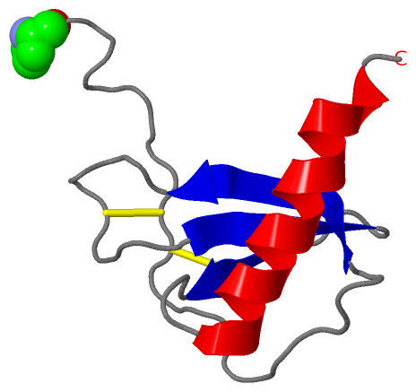
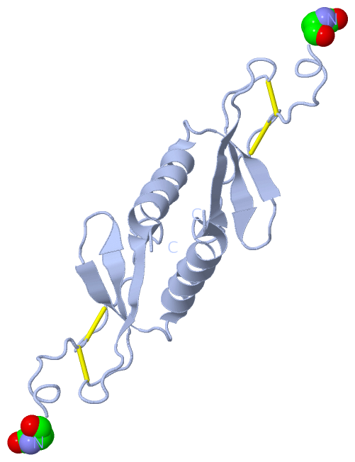
    GO:0008009    chemokine activity    The function of a family of small chemotactic cytokines; their name is derived from their ability to induce directed chemotaxis in nearby responsive cells. All chemokines possess a number of conserved cysteine residues involved in intramolecular disulfide bond formation. Some chemokines are considered pro-inflammatory and can be induced during an immune response to recruit cells of the immune system to a site of infection, while others are considered homeostatic and are involved in controlling the migration of cells during normal processes of tissue maintenance or development. Chemokines are found in all vertebrates, some viruses and some bacteria.
    GO:0005125    cytokine activity    Functions to control the survival, growth, differentiation and effector function of tissues and cells.
    GO:0008201    heparin binding    Interacting selectively and non-covalently with heparin, any member of a group of glycosaminoglycans found mainly as an intracellular component of mast cells and which consist predominantly of alternating alpha-(1->4)-linked D-galactose and N-acetyl-D-glucosamine-6-sulfate residues.
    GO:0016004    phospholipase activator activity    Increases the activity of a phospholipase, an enzyme that catalyzes of the hydrolysis of a glycerophospholipid.
    GO:0004672    protein kinase activity    Catalysis of the phosphorylation of an amino acid residue in a protein, usually according to the reaction: a protein + ATP = a phosphoprotein + ADP.
biological process
    GO:0007186    G-protein coupled receptor signaling pathway    A series of molecular signals that proceeds with an activated receptor promoting the exchange of GDP for GTP on the alpha-subunit of an associated heterotrimeric G-protein complex. The GTP-bound activated alpha-G-protein then dissociates from the beta- and gamma-subunits to further transmit the signal within the cell. The pathway begins with receptor-ligand interaction, or for basal GPCR signaling the pathway begins with the receptor activating its G protein in the absence of an agonist, and ends with regulation of a downstream cellular process, e.g. transcription. The pathway can start from the plasma membrane, Golgi or nuclear membrane (PMID:24568158 and PMID:16902576).
    GO:0006816    calcium ion transport    The directed movement of calcium (Ca) ions into, out of or within a cell, or between cells, by means of some agent such as a transporter or pore.
    GO:0007267    cell-cell signaling    Any process that mediates the transfer of information from one cell to another. This process includes signal transduction in the receiving cell and, where applicable, release of a ligand and any processes that actively facilitate its transport and presentation to the receiving cell. Examples include signaling via soluble ligands, via cell adhesion molecules and via gap junctions.
    GO:0006874    cellular calcium ion homeostasis    Any process involved in the maintenance of an internal steady state of calcium ions at the level of a cell.
    GO:0071346    cellular response to interferon-gamma    Any process that results in a change in state or activity of a cell (in terms of movement, secretion, enzyme production, gene expression, etc.) as a result of an interferon-gamma stimulus. Interferon gamma is the only member of the type II interferon found so far.
    GO:0071347    cellular response to interleukin-1    Any process that results in a change in state or activity of a cell (in terms of movement, secretion, enzyme production, gene expression, etc.) as a result of an interleukin-1 stimulus.
    GO:0071356    cellular response to tumor necrosis factor    Any process that results in a change in state or activity of a cell (in terms of movement, secretion, enzyme production, gene expression, etc.) as a result of a tumor necrosis factor stimulus.
    GO:0070098    chemokine-mediated signaling pathway    A series of molecular signals initiated by the binding of a chemokine to a receptor on the surface of a cell, and ending with regulation of a downstream cellular process, e.g. transcription.
    GO:0006935    chemotaxis    The directed movement of a motile cell or organism, or the directed growth of a cell guided by a specific chemical concentration gradient. Movement may be towards a higher concentration (positive chemotaxis) or towards a lower concentration (negative chemotaxis).
    GO:0006887    exocytosis    A process of secretion by a cell that results in the release of intracellular molecules (e.g. hormones, matrix proteins) contained within a membrane-bounded vesicle. Exocytosis can occur either by full fusion, when the vesicle collapses into the plasma membrane, or by a kiss-and-run mechanism that involves the formation of a transient contact, a pore, between a granule (for exemple of chromaffin cells) and the plasma membrane. The latter process most of the time leads to only partial secretion of the granule content. Exocytosis begins with steps that prepare vesicles for fusion with the membrane (tethering and docking) and ends when molecules are secreted from the cell.
    GO:0006955    immune response    Any immune system process that functions in the calibrated response of an organism to a potential internal or invasive threat.
    GO:0006954    inflammatory response    The immediate defensive reaction (by vertebrate tissue) to infection or injury caused by chemical or physical agents. The process is characterized by local vasodilation, extravasation of plasma into intercellular spaces and accumulation of white blood cells and macrophages.
    GO:0048247    lymphocyte chemotaxis    The directed movement of a lymphocyte in response to an external stimulus.
    GO:0002548    monocyte chemotaxis    The movement of a monocyte in response to an external stimulus.
    GO:0070664    negative regulation of leukocyte proliferation    Any process that stops, prevents, or reduces the frequency, rate or extent of leukocyte proliferation.
    GO:0030593    neutrophil chemotaxis    The directed movement of a neutrophil cell, the most numerous polymorphonuclear leukocyte found in the blood, in response to an external stimulus, usually an infection or wounding.
    GO:0070374    positive regulation of ERK1 and ERK2 cascade    Any process that activates or increases the frequency, rate or extent of signal transduction mediated by the ERK1 and ERK2 cascade.
    GO:0043547    positive regulation of GTPase activity    Any process that activates or increases the activity of a GTPase.
    GO:0050729    positive regulation of inflammatory response    Any process that activates or increases the frequency, rate or extent of the inflammatory response.
    GO:0002687    positive regulation of leukocyte migration    Any process that activates or increases the frequency, rate, or extent of leukocyte migration.
    GO:0006468    protein phosphorylation    The process of introducing a phosphate group on to a protein.
    GO:0009615    response to virus    Any process that results in a change in state or activity of a cell or an organism (in terms of movement, secretion, enzyme production, gene expression, etc.) as a result of a stimulus from a virus.
    GO:0007165    signal transduction    The cellular process in which a signal is conveyed to trigger a change in the activity or state of a cell. Signal transduction begins with reception of a signal (e.g. a ligand binding to a receptor or receptor activation by a stimulus such as light), or for signal transduction in the absence of ligand, signal-withdrawal or the activity of a constitutively active receptor. Signal transduction ends with regulation of a downstream cellular process, e.g. regulation of transcription or regulation of a metabolic process. Signal transduction covers signaling from receptors located on the surface of the cell and signaling via molecules located within the cell. For signaling between cells, signal transduction is restricted to events at and within the receiving cell.
cellular component
    GO:0005623    cell    The basic structural and functional unit of all organisms. Includes the plasma membrane and any external encapsulating structures such as the cell wall and cell envelope.
    GO:0005576    extracellular region    The space external to the outermost structure of a cell. For cells without external protective or external encapsulating structures this refers to space outside of the plasma membrane. This term covers the host cell environment outside an intracellular parasite.
    GO:0005615    extracellular space    That part of a multicellular organism outside the cells proper, usually taken to be outside the plasma membranes, and occupied by fluid.

 Visualization

(-) Interactive Views

Asymmetric Unit
  Complete Structure
    Jena3D(integrated viewing of ligand, site, SAP, PROSITE, SCOP information)
    WebMol | AstexViewer[tm]@PDBe
(Java Applets, require no local installation except for Java; loading may be slow)
    STRAP
(Java WebStart application, automatic local installation, requires Java; full application with system access!)
    RasMol
(require local installation)
    Molscript (VRML)
(requires installation of a VRML viewer; select preferred view via VRML and generate a mono or stereo PDF format file)
 
  Ligands, Modified Residues, Ions
    PCA  [ RasMol | Jena3D ]  +environment [ RasMol | Jena3D ]
 
  Sites
(no "Sites" information available for 1esr)
 
  Cis Peptide Bonds
    Lys A:75 - Pro A:76   [ RasMol ]  
 
Biological Unit
  Complete Structure
    Biological Unit 1  [ Jena3D ]

(-) Still Images

Jmol
  protein: cartoon or spacefill or dots and stick; nucleic acid: cartoon and stick; ligands: spacefill; active site: stick
Molscript
  protein, nucleic acid: cartoon; ligands: spacefill; active site: ball and stick

 Databases and Analysis Tools

(-) Databases

Access by PDB/NDB ID
  1esr
    Family and Domain InformationProDom | SYSTERS
    General Structural InformationGlycoscienceDB | MMDB | NDB | OCA | PDB | PDBe | PDBj | PDBsum | PDBWiki | PQS | PROTEOPEDIA
    Orientation in MembranesOPM
    Protein SurfaceSURFACE
    Secondary StructureDSSP (structure derived) | HSSP (homology derived)
    Structural GenomicsGeneCensus
    Structural NeighboursCE | VAST
    Structure ClassificationCATH | Dali | SCOP
    Validation and Original DataBMRB Data View | BMRB Restraints Grid | EDS | PROCHECK | RECOORD | WHAT_CHECK
 
Access by UniProt ID/Accession number
  CCL8_HUMAN | P80075
    Comparative Protein Structure ModelsModBase
    Genomic InformationEnsembl
    Protein-protein InteractionDIP
    Sequence, Family and Domain InformationInterPro | Pfam | SMART | UniProtKB/SwissProt
 
Access by Enzyme Classificator   (EC Number)
  (no 'Enzyme Classificator' available)
    General Enzyme InformationBRENDA | EC-PDB | Enzyme | IntEnz
    PathwayKEGG | MetaCyc
 
Access by Disease Identifier   (MIM ID)
  (no 'MIM ID' available)
    Disease InformationOMIM
 
Access by GenAge ID
  (no 'GenAge ID' available)
    Age Related InformationGenAge

(-) Analysis Tools

Access by PDB/NDB ID
    Domain InformationXDom
    Interatomic Contacts of Structural UnitsCSU
    Ligand-protein ContactsLPC
    Protein CavitiescastP
    Sequence and Secondary StructurePDBCartoon
    Structure AlignmentSTRAP(Java WebStart application, automatic local installation, requires Java; full application with system access!)
    Structure and Sequence BrowserSTING
 
Access by UniProt ID/Accession number
  CCL8_HUMAN | P80075
    Protein Disorder PredictionDisEMBL | FoldIndex | GLOBPLOT (for more information see DisProt)

 Related Entries

(-) Entries Sharing at Least One Protein Chain (UniProt ID)

(no "Entries Sharing at Least One Protein Chain" available for 1ESR)

(-) Related Entries Specified in the PDB File

1bo0 1BO0 IS THE NMR MINIMIZED AVERAGE STRUCTURE OF HUMAN MCP-3
1dok 1DOK IS THE CRYSTAL STRUCTURE OF HUMAN MCP-1, P-FORM
1dol 1DOL IS THE CRYSTAL STRUCTURE OF HUMAN MCP-1, I-FORM
1dom 1DOM IS THE NMR MINIMIZED AVERAGE STRUCTURE OF HUMAN MCP-1