|
|
|
|
 Description
|
|
Compounds
|
||||||||||||||||||||||||
Chains, Units
Summary Information (see also Sequences/Alignments below) |
Ligands, Modified Residues, Ions (8, 13)
Asymmetric/Biological Unit (8, 13)
|
Sites (3, 3)
Asymmetric Unit (3, 3)
|
SS Bonds (0, 0)| (no "SS Bond" information available for 1E9W) |
Cis Peptide Bonds (0, 0)| (no "Cis Peptide Bond" information available for 1E9W) |
SAPs(SNPs)/Variants (0, 0)| (no "SAP(SNP)/Variant" information available for 1E9W) |
PROSITE Motifs (0, 0)| (no "PROSITE Motif" information available for 1E9W) |
Exons (0, 0)| (no "Exon" information available for 1E9W) |
Sequences/Alignments
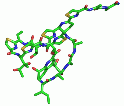
Asymmetric/Biological UnitChain A from PDB Type:PROTEIN Length:19 aligned with THCL_STRAJ | P0C8P8 from UniProtKB/Swiss-Prot Length:72 Alignment length:19 72 64 | THCL_STRAJ 55 MIASASCTTCICTCSCSS- - SCOP domains ------------------- SCOP domains CATH domains ------------------- CATH domains Pfam domains ------------------- Pfam domains SAPs(SNPs) ------------------- SAPs(SNPs) PROSITE ------------------- PROSITE Transcript ------------------- Transcript 1e9w A 0 xIAsAScTtxicTcscssx 18 | | | |9|| |||||| | | | |||| 14-MH6 0-QUA | |||| |15-BB9 3-DHA|||| | 16-DHA 6-BB9| | 17-DHA 8-DBU| 18-NH2 9-DCY 10-TS9 11-BB9 13-BB9
|
||||||||||||||||||||
SCOP Domains (0, 0)| (no "SCOP Domain" information available for 1E9W) |
CATH Domains (0, 0)| (no "CATH Domain" information available for 1E9W) |
Pfam Domains (0, 0)| (no "Pfam Domain" information available for 1E9W) |
Gene Ontology (2, 2)|
Asymmetric/Biological Unit(hide GO term definitions) Chain A (THCL_STRAJ | P0C8P8)
|
||||||||||||||||||||||||
Interactive Views
|
|||||||||||||||||||||||||||||||||||||||||||||||||||||||||||||||||||||||||||||||||||||||||||||||||||||||||||||||||||||||||||||||||||||||||||||||||||||||||||||||||||||||||||||||||||||
Still Images
|
||||||||||||||||
Databases
|
||||||||||||||||||||||||||||||||||||||||||||||||||||||||||||||||||||||||||||||||||||||||||||||||||||||||||||||||||||||||||||||||||||||||||||||||||||||||||||||||
Analysis Tools
|
|||||||||||||||||||||||||||||||||||||||||||||||||||||||||||||
Entries Sharing at Least One Protein Chain (UniProt ID)
Related Entries Specified in the PDB File
|
|