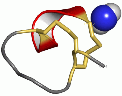
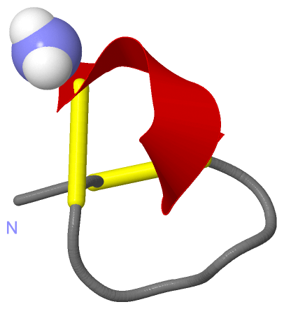
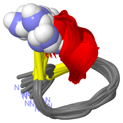
Show PDB file:   
         Plain Text   HTML   (compressed file size)
QuickSearch:   
by PDB,NDB,UniProt,PROSITE Code or Search Term(s)  
(-)NMR Structure - manually
(-)NMR Structure - model 1
(-)NMR Structure - all models
collapse expand < >
Image NMR Structure - manually
NMR Structure - manually  (Jmol Viewer)
Image NMR Structure - model 1
NMR Structure - model 1  (Jmol Viewer)
Image NMR Structure - all models
NMR Structure - all models  (Jmol Viewer)

(-) Description

Title :  NMR SOLUTION STRUCTURE OF ALPHA-CONOTOXIN IM1 POINT MUTATION VARIANT R7L
 
Authors :  J. P. Rogers, P. Luginbuhl, K. Pemberton, P. Harty, D. E. Wemmer, R. C. St
Date :  24 Aug 00  (Deposition) - 27 Dec 00  (Release) - 14 Jun 17  (Revision)
Method :  SOLUTION NMR
Resolution :  NOT APPLICABLE
Chains :  NMR Structure  :  A  (20x)
NMR Structure *:  A  (1x)
Keywords :  Peptide Toxin, Neurotoxin, Neuronal Nicotinic Acetylcholine Receptor Antagonist, Alpha-Conotoxin, Nmr Solution Structure (Keyword Search: [Gene Ontology, PubMed, Web (Google))
 
Reference :  J. P. Rogers, P. Luginbuhl, K. Pemberton, P. Harty, D. E. Wemmer, R. C. Stevens
Structure-Activity Relationships In A Peptidic Alpha7 Nicotinic Acetylcholine Receptor Antagonist
J. Mol. Biol. V. 304 911 2000
PubMed-ID: 11124036  |  Reference-DOI: 10.1006/JMBI.2000.4247
(for further references see the PDB file header)

(-) Compounds

Molecule 1 - ALPHA-CONOTOXIN IM1(R7L)
    ChainsA
    EngineeredYES
    MutationYES
    Organism ScientificCONUS IMPERIALIS
    Organism Taxid35631
    Other DetailsSYNTHESIZED USING STANDARD FMOC CHEMISTRY. IM1 SEQUENCE FOUND NATURALLY IN CONUS IMPERIALIS VENOM
    SyntheticYES

 Structural Features

(-) Chains, Units

  1
NMR Structure (20x)A
NMR Structure * (1x)A

Summary Information (see also Sequences/Alignments below)

(-) Ligands, Modified Residues, Ions  (1, 1)

NMR Structure (1, 1)
No.NameCountTypeFull Name
1NH21Mod. Amino AcidAMINO GROUP
NMR Structure * (1, 1)
No.NameCountTypeFull Name
1NH21Mod. Amino AcidAMINO GROUP

(-) Sites  (0, 0)

(no "Site" information available for 1E75)

(-) SS Bonds  (2, 2)

NMR Structure
No.Residues
1A:2 -A:8
2A:3 -A:12

(-) Cis Peptide Bonds  (0, 0)

(no "Cis Peptide Bond" information available for 1E75)

 Sequence-Structure Mapping

(-) SAPs(SNPs)/Variants  (0, 0)

(no "SAP(SNP)/Variant" information available for 1E75)

(-) PROSITE Motifs  (1, 1)

NMR Structure (1, 1)
 PROSITEUniProtKBPDB
No.IDACDescriptionIDLocationCountLocation
1ALPHA_CONOTOXINPS60014 Alpha-conotoxin family signature.CA1_CONIM6-16  1A:2-12
NMR Structure * (1, 1)
 PROSITEUniProtKBPDB
No.IDACDescriptionIDLocationCountLocation
1ALPHA_CONOTOXINPS60014 Alpha-conotoxin family signature.CA1_CONIM6-16  1A:2-12

(-) Exons   (0, 0)

(no "Exon" information available for 1E75)

(-) Sequences/Alignments

NMR Structure
   Reformat: Number of residues per line =  ('0' or empty: single-line sequence representation)
  Number of residues per labelling interval =   
  UniProt sequence: complete  aligned part    
   Show mapping: SCOP domains CATH domains Pfam domains Secondary structure (by author)
SAPs(SNPs) PROSITE motifs Exons
(details for a mapped element are shown in a popup box when the mouse pointer rests over it)
Chain A from PDB  Type:PROTEIN  Length:13
 aligned with CA1_CONIM | P50983 from UniProtKB/Swiss-Prot  Length:17

    Alignment length:13
                                    14   
             CA1_CONIM    5 GCCSDPRCAWRCG 17
               SCOP domains d1e75a_ A:    SCOP domains
               CATH domains ------------- CATH domains
               Pfam domains ------------- Pfam domains
         Sec.struct. author .......hhhhh. Sec.struct. author
                 SAPs(SNPs) ------------- SAPs(SNPs)
                    PROSITE -ALPHA_CONOT- PROSITE
                 Transcript ------------- Transcript
                  1e75 A  1 GCCSDPLCAWRCx 13
                                    10  |
                                       13-NH2

   Legend:   → Mismatch (orange background)
  - → Gap (green background, '-', border residues have a numbering label)
    → Modified Residue (blue background, lower-case, 'x' indicates undefined single-letter code, labelled with number + name)
  x → Chemical Group (purple background, 'x', labelled with number + name, e.g. ACE or NH2)
  extra numbering lines below/above indicate numbering irregularities and modified residue names etc., number ends below/above '|'

 Classification and Annotation

(-) SCOP Domains  (1, 1)

NMR Structure
(-)
Class: Peptides (792)

(-) CATH Domains  (0, 0)

(no "CATH Domain" information available for 1E75)

(-) Pfam Domains  (0, 0)

(no "Pfam Domain" information available for 1E75)

(-) Gene Ontology  (2, 2)

NMR Structure(hide GO term definitions)
Chain A   (CA1_CONIM | P50983)
cellular component
    GO:0005576    extracellular region    The space external to the outermost structure of a cell. For cells without external protective or external encapsulating structures this refers to space outside of the plasma membrane. This term covers the host cell environment outside an intracellular parasite.
    GO:0035792    other organism postsynaptic membrane    A postsynaptic membrane that is part of another organism, i.e. a secondary organism with which the first organism is interacting. A postsynaptic membrane is a specialized area of membrane facing the presynaptic membrane on the tip of the nerve ending and separated from it by a minute cleft (the synaptic cleft). Neurotransmitters transmit the signal across the synaptic cleft to the postsynaptic membrane.

 Visualization

(-) Interactive Views

NMR Structure
  Complete Structure
    Jena3D(integrated viewing of ligand, site, SAP, PROSITE, SCOP information)
    WebMol | AstexViewer[tm]@PDBe
(Java Applets, require no local installation except for Java; loading may be slow)
    STRAP
(Java WebStart application, automatic local installation, requires Java; full application with system access!)
    RasMol
(require local installation)
    Molscript (VRML)
(requires installation of a VRML viewer; select preferred view via VRML and generate a mono or stereo PDF format file)
 
  Ligands, Modified Residues, Ions
    NH2  [ RasMol | Jena3D ]  +environment [ RasMol | Jena3D ]
 
  Sites
(no "Sites" information available for 1e75)
 
  Cis Peptide Bonds
(no "Cis Peptide Bonds" information available for 1e75)
 

(-) Still Images

Jmol
  protein: cartoon or spacefill or dots and stick; nucleic acid: cartoon and stick; ligands: spacefill; active site: stick
Molscript
  protein, nucleic acid: cartoon; ligands: spacefill; active site: ball and stick

 Databases and Analysis Tools

(-) Databases

Access by PDB/NDB ID
  1e75
    Family and Domain InformationProDom | SYSTERS
    General Structural InformationGlycoscienceDB | MMDB | NDB | OCA | PDB | PDBe | PDBj | PDBsum | PDBWiki | PQS | PROTEOPEDIA
    Orientation in MembranesOPM
    Protein SurfaceSURFACE
    Secondary StructureDSSP (structure derived) | HSSP (homology derived)
    Structural GenomicsGeneCensus
    Structural NeighboursCE | VAST
    Structure ClassificationCATH | Dali | SCOP
    Validation and Original DataBMRB Data View | BMRB Restraints Grid | EDS | PROCHECK | RECOORD | WHAT_CHECK
 
Access by UniProt ID/Accession number
  CA1_CONIM | P50983
    Comparative Protein Structure ModelsModBase
    Genomic InformationEnsembl
    Protein-protein InteractionDIP
    Sequence, Family and Domain InformationInterPro | Pfam | SMART | UniProtKB/SwissProt
 
Access by Enzyme Classificator   (EC Number)
  (no 'Enzyme Classificator' available)
    General Enzyme InformationBRENDA | EC-PDB | Enzyme | IntEnz
    PathwayKEGG | MetaCyc
 
Access by Disease Identifier   (MIM ID)
  (no 'MIM ID' available)
    Disease InformationOMIM
 
Access by GenAge ID
  (no 'GenAge ID' available)
    Age Related InformationGenAge

(-) Analysis Tools

Access by PDB/NDB ID
    Domain InformationXDom
    Interatomic Contacts of Structural UnitsCSU
    Ligand-protein ContactsLPC
    Protein CavitiescastP
    Sequence and Secondary StructurePDBCartoon
    Structure AlignmentSTRAP(Java WebStart application, automatic local installation, requires Java; full application with system access!)
    Structure and Sequence BrowserSTING
 
Access by UniProt ID/Accession number
  CA1_CONIM | P50983
    Protein Disorder PredictionDisEMBL | FoldIndex | GLOBPLOT (for more information see DisProt)

 Related Entries

(-) Entries Sharing at Least One Protein Chain (UniProt ID)

UniProtKB/Swiss-Prot
        CA1_CONIM | P509831cnl 1e74 1e76 1g2g 1im1 1imi 2bc7 2bc8 2byp 2c9t 2igu 2moa

(-) Related Entries Specified in the PDB File

4846
1cnl ALPHA-CONOTOXIN IMI (10 STRUCTURES)
1e74 NMR SOLUTION STRUCTURE OF ALPHA-CONOTOXIN IM1 POINT MUTATION VARIANT R11E (20 STRUCTURES)
1e76 NMR SOLUTION STRUCTURE OF ALPHA-CONOTOXIN IM1 POINT MUTATION VARIANT D5N (20 STRUCTURES)
1im1 NMR SOLUTION STRUCTURE OF ALPHA-CONOTOXIN IM1, (20 STRUCTURES)
1imi SOLUTION STRUCTURE OF ALPHA-CONOTOXIN IM1