|
|
|
|
 Description
|
|
Compounds
|
||||||||||||||||||||||||||||||||||||||||||||||||||||||||||||||||||||||||
Chains, Units
Summary Information (see also Sequences/Alignments below) |
Ligands, Modified Residues, Ions (0, 0)| (no "Ligand,Modified Residues,Ions" information available for 1E5U) |
Sites (0, 0)| (no "Site" information available for 1E5U) |
SS Bonds (1, 1)
NMR Structure
|
||||||||
Cis Peptide Bonds (0, 0)| (no "Cis Peptide Bond" information available for 1E5U) |
SAPs(SNPs)/Variants (0, 0)| (no "SAP(SNP)/Variant" information available for 1E5U) |
PROSITE Motifs (0, 0)| (no "PROSITE Motif" information available for 1E5U) |
Exons (0, 0)| (no "Exon" information available for 1E5U) |
Sequences/Alignments
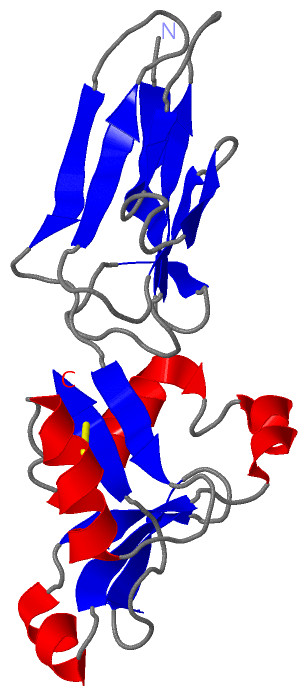
NMR StructureChain I from PDB Type:PROTEIN Length:187 aligned with EAE_ECO27 | P19809 from UniProtKB/Swiss-Prot Length:939 Alignment length:187 762 772 782 792 802 812 822 832 842 852 862 872 882 892 902 912 922 932 EAE_ECO27 753 TLTIDDGNIEIVGTGVKGKLPTVWLQYGQVNLKASGGNGKYTWRSANPAIASVDASSGQVTLKEKGTTTISVISSDNQTATYTIATPNSLIVPNMSKRVTYNDAVNTCKNFGGKLPSSQNELENVFKAWGAANKYEYYKSSQTIISWVQQTAQDAKSGVASTYDLVKQNPLNNIKASESNAYATCVK 939 SCOP domains d1e5ui1 I:1-89 Intimin d1e5ui2 I:90-187 Intimin SCOP domains CATH domains 1e5uI01 I:1-90 [code=2.60.40.1080, no name defined] 1e5uI02 I:91-187 Mannose-Binding Protein A, subunit A CATH domains Pfam domains ------------------------------------------------------------------------------------------------------------------------------------------------------------------------------------------- Pfam domains SAPs(SNPs) ------------------------------------------------------------------------------------------------------------------------------------------------------------------------------------------- SAPs(SNPs) PROSITE ------------------------------------------------------------------------------------------------------------------------------------------------------------------------------------------- PROSITE Transcript ------------------------------------------------------------------------------------------------------------------------------------------------------------------------------------------- Transcript 1e5u I 1 TLTIDDGNIEIVGTGVKGKLPTVWLQYGQVNLKASGGNGKYTWRSANPAIASVDASSGQVTLKEKGTTTISVISSDNQTATYTIATPNSLIVPNMSKRVTYNDAVNTCKNFGGKLPSSQNELENVFKAWGAANKYEYYKSSQTIISWVQQTAQDAKSGVASTYDLVKQNPLNNIKASESNAYATCVK 187 10 20 30 40 50 60 70 80 90 100 110 120 130 140 150 160 170 180
|
||||||||||||||||||||
SCOP Domains (2, 2)
NMR Structure
 |
CATH Domains (2, 2)
NMR Structure
|
Pfam Domains (0, 0)| (no "Pfam Domain" information available for 1E5U) |
Gene Ontology (5, 5)|
NMR Structure(hide GO term definitions) Chain I (EAE_ECO27 | P19809)
|
||||||||||||||||||||||||||||||||||||||||||
Interactive Views
|
||||||||||||||||||||||||||||||||||||||||||||||||||||||||||||||||||||||||||||||||||||||||||||||||||||||||||||||||||||
Still Images
|
||||||||||||||||
Databases
|
||||||||||||||||||||||||||||||||||||||||||||||||||||||||||||||||||||||||||||||||||||||||||||||||||||||||||||||||||||||||||||||||||||||||||||||||||||||||||||||||
Analysis Tools
|
|||||||||||||||||||||||||||||||||||||||||||||||||||||||||||||
Entries Sharing at Least One Protein Chain (UniProt ID)
Related Entries Specified in the PDB File
|
|