|
|
|
|
 Description
|
|
Compounds
|
||||||||||||||||||||||||||||||||||||||||||||
Chains, Units
Summary Information (see also Sequences/Alignments below) |
Ligands, Modified Residues, Ions (5, 14)| Asymmetric Unit (5, 14) Biological Unit 1 (3, 36) |
Sites (13, 13)
Asymmetric Unit (13, 13)
|
SS Bonds (0, 0)| (no "SS Bond" information available for 1E12) |
Cis Peptide Bonds (0, 0)| (no "Cis Peptide Bond" information available for 1E12) |
SAPs(SNPs)/Variants (0, 0)| (no "SAP(SNP)/Variant" information available for 1E12) |
PROSITE Motifs (2, 2)
Asymmetric Unit (2, 2)
|
||||||||||||||||||||||||||||||||||||||||||||||||||||||||||||||||||||||||||||||||
Exons (0, 0)| (no "Exon" information available for 1E12) |
Sequences/Alignments
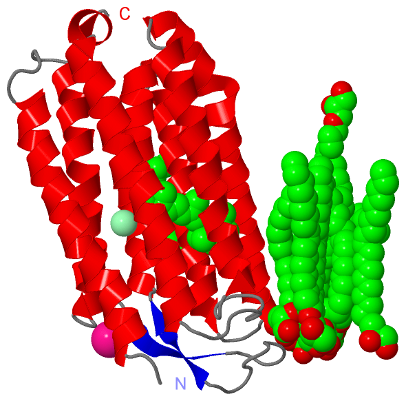
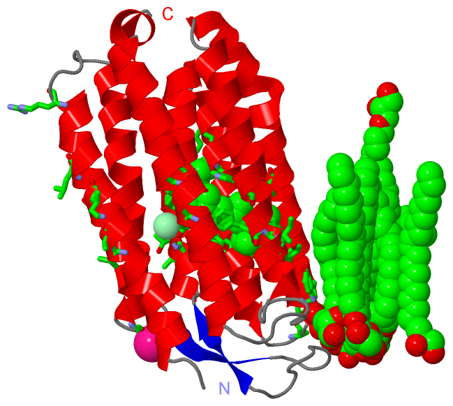
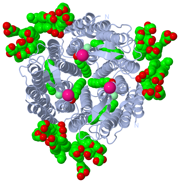
Asymmetric UnitChain A from PDB Type:PROTEIN Length:239 aligned with BACH_HALS3 | B0R2U4 from UniProtKB/Swiss-Prot Length:274 Alignment length:239 33 43 53 63 73 83 93 103 113 123 133 143 153 163 173 183 193 203 213 223 233 243 253 BACH_HALS3 24 RENALLSSSLWVNVALAGIAILVFVYMGRTIRPGRPRLIWGATLMIPLVSISSYLGLLSGLTVGMIEMPAGHALAGEMVRSQWGRYLTWALSTPMILLALGLLADVDLGSLFTVIAADIGMCVTGLAAAMTTSALLFRWAFYAISCAFFVVVLSALVTDWAASASSAGTAEIFDTLRVLTVVLWLGYPIVWAVGVEGLALVQSVGVTSWAYSVLDVFAKYVFAFILLRWVANNERTVAV 262 SCOP domains d1e12a_ A: Halorhodopsin SCOP domains CATH domains 1e12A00 A:24-262 Rhopdopsin 7-helix transmembrane proteins CATH domains Pfam domains ----------------------------------------------------------------------------------------------------------------------------------------------------------------------------------------------------------------------------------------------- Pfam domains SAPs(SNPs) ----------------------------------------------------------------------------------------------------------------------------------------------------------------------------------------------------------------------------------------------- SAPs(SNPs) PROSITE (2) ------------------------------------------------------------------------------------BACTERIAL_OPS-----------------------------------------------------------------------------------------------------------------BACTERIAL_OP----------------- PROSITE (2) Transcript ----------------------------------------------------------------------------------------------------------------------------------------------------------------------------------------------------------------------------------------------- Transcript 1e12 A 24 RENALLSSSLWVNVALAGIAILVFVYMGRTIRPGRPRLIWGATLMIPLVSISSYLGLLSGLTVGMIEMPAGHALAGEMVRSQWGRYLTWALSTPMILLALGLLADVDLGSLFTVIAADIGMCVTGLAAAMTTSALLFRWAFYAISCAFFVVVLSALVTDWAASASSAGTAEIFDTLRVLTVVLWLGYPIVWAVGVEGLALVQSVGATSWAYSVLDVFAKYVFAFILLRWVANNERTVAV 262 33 43 53 63 73 83 93 103 113 123 133 143 153 163 173 183 193 203 213 223 233 243 253 Chain A from PDB Type:PROTEIN Length:239 aligned with BACH_HALSA | P0DMH7 from UniProtKB/Swiss-Prot Length:274 Alignment length:239 33 43 53 63 73 83 93 103 113 123 133 143 153 163 173 183 193 203 213 223 233 243 253 BACH_HALSA 24 RENALLSSSLWVNVALAGIAILVFVYMGRTIRPGRPRLIWGATLMIPLVSISSYLGLLSGLTVGMIEMPAGHALAGEMVRSQWGRYLTWALSTPMILLALGLLADVDLGSLFTVIAADIGMCVTGLAAAMTTSALLFRWAFYAISCAFFVVVLSALVTDWAASASSAGTAEIFDTLRVLTVVLWLGYPIVWAVGVEGLALVQSVGVTSWAYSVLDVFAKYVFAFILLRWVANNERTVAV 262 SCOP domains d1e12a_ A: Halorhodopsin SCOP domains CATH domains 1e12A00 A:24-262 Rhopdopsin 7-helix transmembrane proteins CATH domains Pfam domains ----------------------------------------------------------------------------------------------------------------------------------------------------------------------------------------------------------------------------------------------- Pfam domains SAPs(SNPs) ----------------------------------------------------------------------------------------------------------------------------------------------------------------------------------------------------------------------------------------------- SAPs(SNPs) PROSITE ------------------------------------------------------------------------------------BACTERIAL_OPS-----------------------------------------------------------------------------------------------------------------BACTERIAL_OP----------------- PROSITE Transcript ----------------------------------------------------------------------------------------------------------------------------------------------------------------------------------------------------------------------------------------------- Transcript 1e12 A 24 RENALLSSSLWVNVALAGIAILVFVYMGRTIRPGRPRLIWGATLMIPLVSISSYLGLLSGLTVGMIEMPAGHALAGEMVRSQWGRYLTWALSTPMILLALGLLADVDLGSLFTVIAADIGMCVTGLAAAMTTSALLFRWAFYAISCAFFVVVLSALVTDWAASASSAGTAEIFDTLRVLTVVLWLGYPIVWAVGVEGLALVQSVGATSWAYSVLDVFAKYVFAFILLRWVANNERTVAV 262 33 43 53 63 73 83 93 103 113 123 133 143 153 163 173 183 193 203 213 223 233 243 253
|
||||||||||||||||||||
SCOP Domains (1, 1)
Asymmetric Unit
|
CATH Domains (1, 1)
Asymmetric Unit
 |
Pfam Domains (0, 0)| (no "Pfam Domain" information available for 1E12) |
Gene Ontology (11, 22)|
Asymmetric Unit(hide GO term definitions) Chain A (BACH_HALS3 | B0R2U4)
Chain A (BACH_HALSA | P0DMH7)
|
||||||||||||||||||||||||||||||||||||||||||||||||||||||||||||||||||||||||||||||||||||||||||||||||||||||||||||||||||||||||||||||||||||||||||||||||||||||||||||||||||||||||
Interactive Views
|
||||||||||||||||||||||||||||||||||||||||||||||||||||||||||||||||||||||||||||||||||||||||||||||||||||||||||||||||||||||||||||||||||||||||||||||||||||||||||||||||||||||||||||||||||||||||||||||||||||||||||||||||||||||||||||||||||||||||||||||||||||||||
Still Images
|
||||||||||||||||
Databases
|
||||||||||||||||||||||||||||||||||||||||||||||||||||||||||||||||||||||||||||||||||||||||||||||||||||||||||||||||||||||||||||||||||||||||||||||||||||||||||||||||||||||||||||||||||||||||||
Analysis Tools
|
||||||||||||||||||||||||||||||||||||||||||||||||||||||||||||||||||||||||
Entries Sharing at Least One Protein Chain (UniProt ID)
Related Entries Specified in the PDB File
|
|