Show PDB file:   
         Plain Text   HTML   (compressed file size)
QuickSearch:   
by PDB,NDB,UniProt,PROSITE Code or Search Term(s)  
(-)Biol.Unit 1 - manually
(-)Asym.Unit - manually
(-)Asymmetric Unit
(-)Asym. Unit - sites
(-)Biological Unit 1
(-)Biol. Unit 1 - sites
collapse expand < >
Image Biol.Unit 1 - manually
Biol.Unit 1 - manually  (Jmol Viewer)
Image Asym.Unit - manually
Asym.Unit - manually  (Jmol Viewer)
Image Asymmetric Unit
Asymmetric Unit  (Jmol Viewer)
Image Asym. Unit - sites
Asym. Unit - sites  (Jmol Viewer)
Image Biological Unit 1
Biological Unit 1  (Jmol Viewer)
Image Biol. Unit 1 - sites
Biol. Unit 1 - sites  (Jmol Viewer)

(-) Description

Title :  STRUCTURE OF THE FIBER-FORMING PROTEIN PILIN AT 2.6 ANGSTROMS RESOLUTION
 
Authors :  K. T. Forest, H. E. Parge, J. A. Tainer
Date :  13 Nov 97  (Deposition) - 29 Apr 98  (Release) - 13 Jul 11  (Revision)
Method :  X-RAY DIFFRACTION
Resolution :  2.60
Chains :  Asym. Unit :  A
Biol. Unit 1:  A  (2x)
Keywords :  Type Iv Pilin, Fiber-Forming Protein, Membrane Protein, Dna Inding Protein, Contractile Protein, Cell Adhesion (Keyword Search: [Gene Ontology, PubMed, Web (Google))
 
Reference :  H. E. Parge, K. T. Forest, M. J. Hickey, D. A. Christensen, E. D. Getzoff J. A. Tainer
Structure Of The Fibre-Forming Protein Pilin At 2. 6 A Resolution.
Nature V. 378 32 1995
PubMed-ID: 7477282  |  Reference-DOI: 10.1038/378032A0

(-) Compounds

Molecule 1 - TYPE 4 PILIN
    Cellular LocationEXTRACELLULAR
    ChainsA
    OrganellePILUS
    Organism ScientificNEISSERIA GONORRHOEAE
    Organism Taxid485
    StrainMS11
    SynonymFIMBRIAE

 Structural Features

(-) Chains, Units

  1
Asymmetric Unit A
Biological Unit 1 (2x)A

Summary Information (see also Sequences/Alignments below)
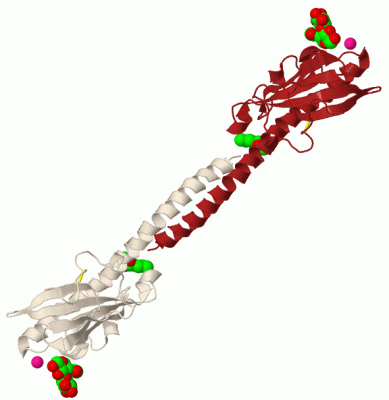
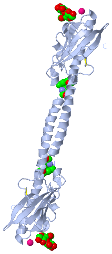
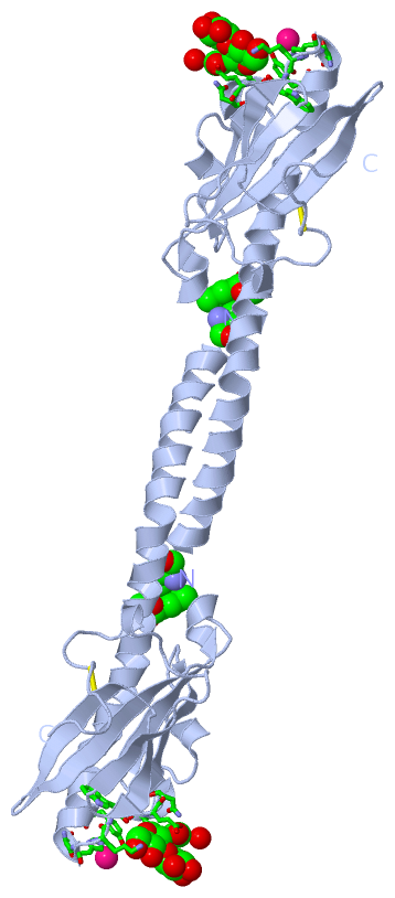
(-) Ligands, Modified Residues, Ions  (5, 5)

Asymmetric Unit (5, 5)
No.NameCountTypeFull Name
10A91Mod. Amino AcidMETHYL L-PHENYLALANINATE
2GLA1Ligand/IonALPHA D-GALACTOSE
3HTO1Ligand/IonHEPTANE-1,2,3-TRIOL
4NAG1Ligand/IonN-ACETYL-D-GLUCOSAMINE
5PT1Ligand/IonPLATINUM (II) ION
Biological Unit 1 (4, 8)
No.NameCountTypeFull Name
10A92Mod. Amino AcidMETHYL L-PHENYLALANINATE
2GLA2Ligand/IonALPHA D-GALACTOSE
3HTO2Ligand/IonHEPTANE-1,2,3-TRIOL
4NAG2Ligand/IonN-ACETYL-D-GLUCOSAMINE
5PT-1Ligand/IonPLATINUM (II) ION

(-) Sites  (4, 4)

Asymmetric Unit (4, 4)
No.NameEvidenceResiduesDescription
1AC1SOFTWARENAG A:161 , HOH A:387BINDING SITE FOR RESIDUE GLA A 160
2AC2SOFTWARETYR A:50 , ASN A:53 , HIS A:54 , LYS A:56 , TRP A:57 , GLU A:59 , ASN A:60 , SER A:63 , GLA A:160BINDING SITE FOR RESIDUE NAG A 161
3AC3SOFTWAREHIS A:54 , HOH A:333 , HOH A:348BINDING SITE FOR RESIDUE PT A 200
4AC4SOFTWARE0A9 A:1BINDING SITE FOR RESIDUE HTO A 162

(-) SS Bonds  (1, 1)

Asymmetric Unit
No.Residues
1A:121 -A:151

(-) Cis Peptide Bonds  (0, 0)

(no "Cis Peptide Bond" information available for 1AY2)

 Sequence-Structure Mapping

(-) SAPs(SNPs)/Variants  (0, 0)

(no "SAP(SNP)/Variant" information available for 1AY2)

(-) PROSITE Motifs  (0, 0)

(no "PROSITE Motif" information available for 1AY2)

(-) Exons   (0, 0)

(no "Exon" information available for 1AY2)

(-) Sequences/Alignments

Asymmetric Unit
   Reformat: Number of residues per line =  ('0' or empty: single-line sequence representation)
  Number of residues per labelling interval =   
  UniProt sequence: complete  aligned part    
   Show mapping: SCOP domains CATH domains Pfam domains Secondary structure (by author)
SAPs(SNPs) PROSITE motifs Exons
(details for a mapped element are shown in a popup box when the mouse pointer rests over it)
Chain A from PDB  Type:PROTEIN  Length:158
 aligned with FMM1_NEIGO | P02974 from UniProtKB/Swiss-Prot  Length:165

    Alignment length:158
                                    17        27        37        47        57        67        77        87        97       107       117       127       137       147       157        
           FMM1_NEIGO     8 FTLIELMIVIAIVGILAAVALPAYQDYTARAQVSEAILLAEGQKSAVTEYYLNHGKWPENNTSAGVASPPSDIKGKYVKEVEVKNGVVTATMLSSGVNNEIKGKKLSLWARRENGSVKWFCGQPVTRTDDDTVADAKDGKEIDTKHLPSTCRDNFDAK 165
               SCOP domains d1ay2a_ A: Pilin Gc                                                                                                                                            SCOP domains
               CATH domains -1ay2A00 A:2-158 Glycoprotein, Type 4 Pilin                                                                                                                    CATH domains
               Pfam domains -------------------------------------------------------------------------------------------------------------------------------------------------------------- Pfam domains
         Sec.struct. author .hhhhhhhhhhhhhhhhhhhhhhhhhhhhhhhhhhhhhhhhhhhhhhhhhhhhh.....hhhhh....hhhh.....eeeeeee..eeeeee......hhhh..eeeeeeeee..eeeeeeee.eeeee..eeee........hhh............ Sec.struct. author
                 SAPs(SNPs) -------------------------------------------------------------------------------------------------------------------------------------------------------------- SAPs(SNPs)
                    PROSITE -------------------------------------------------------------------------------------------------------------------------------------------------------------- PROSITE
                 Transcript -------------------------------------------------------------------------------------------------------------------------------------------------------------- Transcript
                 1ay2 A   1 fTLIELMIVIAIVGILAAVALPAYQDYTARAQVSEAILLAEGQKSAVTEYYLNHGKWPENNTSAGVASPPSDIKGKYVKEVEVKNGVVTATMLSSGVNNEIKGKKLSLWARRENGSVKWFCGQPVTRTDDDTVADAKDGKEIDTKHLPSTCRDNFDAK 158
                            |       10        20        30        40        50        60        70        80        90       100       110       120       130       140       150        
                            |                                                                                                                                                             
                            1-0A9                                                                                                                                                         

   Legend:   → Mismatch (orange background)
  - → Gap (green background, '-', border residues have a numbering label)
    → Modified Residue (blue background, lower-case, 'x' indicates undefined single-letter code, labelled with number + name)
  x → Chemical Group (purple background, 'x', labelled with number + name, e.g. ACE or NH2)
  extra numbering lines below/above indicate numbering irregularities and modified residue names etc., number ends below/above '|'

 Classification and Annotation

(-) SCOP Domains  (1, 1)

Asymmetric Unit

(-) CATH Domains  (1, 1)

Asymmetric Unit
(-)
Class: Alpha Beta (26913)

(-) Pfam Domains  (0, 0)

(no "Pfam Domain" information available for 1AY2)

(-) Gene Ontology  (2, 2)

Asymmetric Unit(hide GO term definitions)
Chain A   (FMM1_NEIGO | P02974)
biological process
    GO:0007155    cell adhesion    The attachment of a cell, either to another cell or to an underlying substrate such as the extracellular matrix, via cell adhesion molecules.
cellular component
    GO:0009289    pilus    A proteinaceous hair-like appendage on the surface of bacteria ranging from 2-8 nm in diameter.

 Visualization

(-) Interactive Views

Asymmetric Unit
  Complete Structure
    Jena3D(integrated viewing of ligand, site, SAP, PROSITE, SCOP information)
    WebMol | AstexViewer[tm]@PDBe
(Java Applets, require no local installation except for Java; loading may be slow)
    STRAP
(Java WebStart application, automatic local installation, requires Java; full application with system access!)
    RasMol
(require local installation)
    Molscript (VRML)
(requires installation of a VRML viewer; select preferred view via VRML and generate a mono or stereo PDF format file)
 
  Ligands, Modified Residues, Ions
    0A9  [ RasMol | Jena3D ]  +environment [ RasMol | Jena3D ]
    GLA  [ RasMol | Jena3D ]  +environment [ RasMol | Jena3D ]
    HTO  [ RasMol | Jena3D ]  +environment [ RasMol | Jena3D ]
    NAG  [ RasMol | Jena3D ]  +environment [ RasMol | Jena3D ]
    PT  [ RasMol | Jena3D ]  +environment [ RasMol | Jena3D ]
 
  Sites
    AC1  [ RasMol ]  +environment [ RasMol ]
    AC2  [ RasMol ]  +environment [ RasMol ]
    AC3  [ RasMol ]  +environment [ RasMol ]
    AC4  [ RasMol ]  +environment [ RasMol ]
 
  Cis Peptide Bonds
(no "Cis Peptide Bonds" information available for 1ay2)
 
Biological Unit
  Complete Structure
    Biological Unit 1  [ Jena3D ]

(-) Still Images

Jmol
  protein: cartoon or spacefill or dots and stick; nucleic acid: cartoon and stick; ligands: spacefill; active site: stick
Molscript
  protein, nucleic acid: cartoon; ligands: spacefill; active site: ball and stick

 Databases and Analysis Tools

(-) Databases

Access by PDB/NDB ID
  1ay2
    Family and Domain InformationProDom | SYSTERS
    General Structural InformationGlycoscienceDB | MMDB | NDB | OCA | PDB | PDBe | PDBj | PDBsum | PDBWiki | PQS | PROTEOPEDIA
    Orientation in MembranesOPM
    Protein SurfaceSURFACE
    Secondary StructureDSSP (structure derived) | HSSP (homology derived)
    Structural GenomicsGeneCensus
    Structural NeighboursCE | VAST
    Structure ClassificationCATH | Dali | SCOP
    Validation and Original DataBMRB Data View | BMRB Restraints Grid | EDS | PROCHECK | RECOORD | WHAT_CHECK
 
Access by UniProt ID/Accession number
  FMM1_NEIGO | P02974
    Comparative Protein Structure ModelsModBase
    Genomic InformationEnsembl
    Protein-protein InteractionDIP
    Sequence, Family and Domain InformationInterPro | Pfam | SMART | UniProtKB/SwissProt
 
Access by Enzyme Classificator   (EC Number)
  (no 'Enzyme Classificator' available)
    General Enzyme InformationBRENDA | EC-PDB | Enzyme | IntEnz
    PathwayKEGG | MetaCyc
 
Access by Disease Identifier   (MIM ID)
  (no 'MIM ID' available)
    Disease InformationOMIM
 
Access by GenAge ID
  (no 'GenAge ID' available)
    Age Related InformationGenAge

(-) Analysis Tools

Access by PDB/NDB ID
    Domain InformationXDom
    Interatomic Contacts of Structural UnitsCSU
    Ligand-protein ContactsLPC
    Protein CavitiescastP
    Sequence and Secondary StructurePDBCartoon
    Structure AlignmentSTRAP(Java WebStart application, automatic local installation, requires Java; full application with system access!)
    Structure and Sequence BrowserSTING
 
Access by UniProt ID/Accession number
  FMM1_NEIGO | P02974
    Protein Disorder PredictionDisEMBL | FoldIndex | GLOBPLOT (for more information see DisProt)

 Related Entries

(-) Entries Sharing at Least One Protein Chain (UniProt ID)

UniProtKB/Swiss-Prot
        FMM1_NEIGO | P029742hi2 2hil 2pil 5vxx

(-) Related Entries Specified in the PDB File

(no "Related Entries Specified in the PDB File" available for 1AY2)