|
|
|
|
 Description
|
|
Compounds
|
||||||||||||||||||||||||||||||||||||||||||||||||
Chains, Units
Summary Information (see also Sequences/Alignments below) |
Ligands, Modified Residues, Ions (1, 1)
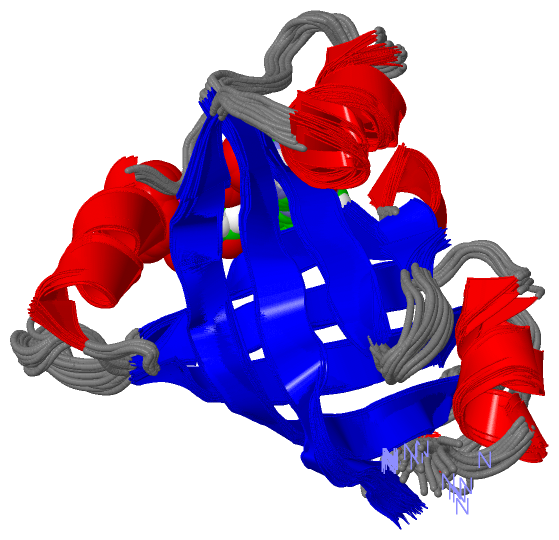
NMR Structure (1, 1)
|
Sites (2, 2)
NMR Structure (2, 2)
|
SS Bonds (0, 0)| (no "SS Bond" information available for 1AXJ) |
Cis Peptide Bonds (0, 0)| (no "Cis Peptide Bond" information available for 1AXJ) |
SAPs(SNPs)/Variants (0, 0)| (no "SAP(SNP)/Variant" information available for 1AXJ) |
PROSITE Motifs (0, 0)| (no "PROSITE Motif" information available for 1AXJ) |
Exons (0, 0)| (no "Exon" information available for 1AXJ) |
Sequences/Alignments
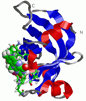
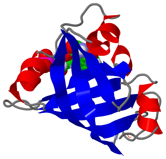
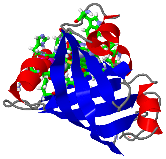
NMR StructureChain A from PDB Type:PROTEIN Length:122 aligned with FMNB_DESVM | Q46604 from UniProtKB/Swiss-Prot Length:122 Alignment length:122 10 20 30 40 50 60 70 80 90 100 110 120 FMNB_DESVM 1 MLPGTFFEVLKNEGVVAIATQGEDGPHLVNTWNSYLKVLDGNRIVVPVGGMHKTEANVARDERVLMTLGSRKVAGRNGPGTGFLIRGSAAFRTDGPEFEAIARFKWARAALVITVVSAEQTL 122 SCOP domains d1axja_ A: FMN-binding protein SCOP domains CATH domains 1axjA00 A:1-122 Electron Transport, Fmn-binding Protein; Chain A CATH domains Pfam domains -------------------------------------------------------------------------------------------------------------------------- Pfam domains SAPs(SNPs) -------------------------------------------------------------------------------------------------------------------------- SAPs(SNPs) PROSITE -------------------------------------------------------------------------------------------------------------------------- PROSITE Transcript -------------------------------------------------------------------------------------------------------------------------- Transcript 1axj A 1 MLPGTFFEVLKNEGVVAIATQGEDGPHLVNTWNSYLKVLDGNRIVVPVGGMHKTEANVARDERVLMTLGSRKVAGRNGPGTGFLIRGSAAFRTDGPEFEAIARFKWARAALVITVVSAEQTL 122 10 20 30 40 50 60 70 80 90 100 110 120
|
||||||||||||||||||||
SCOP Domains (1, 1)
NMR Structure
 |
CATH Domains (1, 1)
NMR Structure
|
Pfam Domains (0, 0)| (no "Pfam Domain" information available for 1AXJ) |
Gene Ontology (6, 6)|
NMR Structure(hide GO term definitions) Chain A (FMNB_DESVM | Q46604)
|
||||||||||||||||||||||||||||||||||||||||||||||||||||||
Interactive Views
|
|||||||||||||||||||||||||||||||||||||||||||||||||||||||||||||||||||||||||||||||||||||||||||||||||||||||||||||||||||||||||||||
Still Images
|
||||||||||||||||
Databases
|
||||||||||||||||||||||||||||||||||||||||||||||||||||||||||||||||||||||||||||||||||||||||||||||||||||||||||||||||||||||||||||||||||||||||||||||||||||||||||||||||
Analysis Tools
|
|||||||||||||||||||||||||||||||||||||||||||||||||||||||||||||
Entries Sharing at Least One Protein Chain (UniProt ID)
Related Entries Specified in the PDB File
|
|