|
|
|
|
 Description
|
|
Compounds
|
||||||||||||||||||||||||||||||||||||||||||
Chains, Units
Summary Information (see also Sequences/Alignments below) |
Ligands, Modified Residues, Ions (5, 36)| Asymmetric/Biological Unit (5, 36) |
Sites (2, 2)
Asymmetric Unit (2, 2)
|
SS Bonds (0, 0)| (no "SS Bond" information available for 1ALX) |
Cis Peptide Bonds (0, 0)| (no "Cis Peptide Bond" information available for 1ALX) |
SAPs(SNPs)/Variants (0, 0)| (no "SAP(SNP)/Variant" information available for 1ALX) |
PROSITE Motifs (0, 0)| (no "PROSITE Motif" information available for 1ALX) |
Exons (0, 0)| (no "Exon" information available for 1ALX) |
Sequences/Alignments
Asymmetric/Biological Unit
Chain A from PDB Type:PROTEIN Length:17
SCOP domains d1alxa_ A: SCOP domains
CATH domains ----------------- CATH domains
Pfam domains ----------------- Pfam domains
SAPs(SNPs) ----------------- SAPs(SNPs)
PROSITE ----------------- PROSITE
Transcript ----------------- Transcript
1alx A 1 xGAxAxVxWxYWxWxWx 16
| | | |10 | | |
| | | | | | | |
1-FVA| | | | | |
4-DLE | | | |
6-DVA | | |
8-DVA| | |
10-DLE| |
12-DLE
14-DLE
16-ETA
Chain B from PDB Type:PROTEIN Length:16
SCOP domains d1alxb_ B: SCOP domains
CATH domains ---------------- CATH domains
Pfam domains ---------------- Pfam domains
SAPs(SNPs) ---------------- SAPs(SNPs)
PROSITE ---------------- PROSITE
Transcript ---------------- Transcript
1alx B 1 xGAxAxVxWxWxWxWx 16
| | | |10 | | |
| | | | | | | |
1-FVA| | | | | |
4-DLE | | | |
6-DVA | | |
8-DVA | |
10-DLE |
12-DLE
14-DLE
16-ETA
|
||||||||||||||||||||
SCOP Domains (1, 2)
Asymmetric/Biological Unit
|
CATH Domains (0, 0)| (no "CATH Domain" information available for 1ALX) |
Pfam Domains (0, 0)| (no "Pfam Domain" information available for 1ALX) |
Gene Ontology (0, 0)|
Asymmetric/Biological Unit(hide GO term definitions)
(no "Gene Ontology" information available for 1ALX)
|
Interactive Views
|
|||||||||||||||||||||||||||||||||||||||||||||||||||||||||||||||||||||||||||||||||||||||||||||||||||||||||||||||||||||||||||||||||||||||||||||||||||||||||
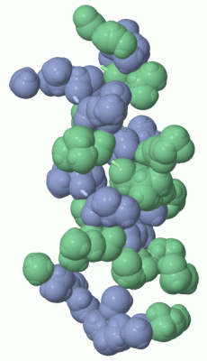
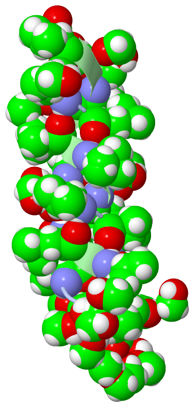
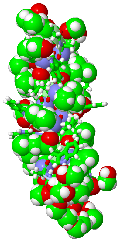
Still Images
|
||||||||||||||||
Databases
|
||||||||||||||||||||||||||||||||||||||||||||||||||||||||||||||||||||||||||||||||||||||||||||||||||||||||||||||||||||||||||||||||||||||||||||||||||||||||||||||||
Analysis Tools
|
|||||||||||||||||||||||||||||||||||||||||||||||||||||||||||||
Entries Sharing at Least One Protein Chain (UniProt ID)
Related Entries Specified in the PDB File
|
|