|
|
|
|
 Description
|
|
Compounds
|
||||||||||||||||||||||||||||||||||||||||||||||||||||
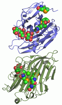
Chains, Units
Summary Information (see also Sequences/Alignments below) |
Ligands, Modified Residues, Ions (3, 16)| Asymmetric/Biological Unit (3, 16) |
Sites (8, 8)
Asymmetric Unit (8, 8)
|
SS Bonds (0, 0)| (no "SS Bond" information available for 1ZM1) |
Cis Peptide Bonds (4, 4)
Asymmetric/Biological Unit
|
||||||||||||||||||||
SAPs(SNPs)/Variants (0, 0)| (no "SAP(SNP)/Variant" information available for 1ZM1) |
PROSITE Motifs (1, 2)| Asymmetric/Biological Unit (1, 2) |
Exons (0, 0)| (no "Exon" information available for 1ZM1) |
Sequences/Alignments
Asymmetric/Biological UnitChain A from PDB Type:PROTEIN Length:241 aligned with GUB_FIBSS | P17989 from UniProtKB/Swiss-Prot Length:349 Alignment length:241 36 46 56 66 76 86 96 106 116 126 136 146 156 166 176 186 196 206 216 226 236 246 256 266 GUB_FIBSS 27 AKDFSGAELYTLEEVQYGKFEARMKMAAASGTVSSMFLYQNGSEIADGRPWVEVDIEVLGKNPGSFQSNIITGKAGAQKTSEKHHAVSPAADQAFHTYGLEWTPNYVRWTVDGQEVRKTEGGQVSNLTGTQGLRFNLWSSESAAWVGQFDESKLPLFQFINWVKVYKYTPGQGEGGSDFTLDWTDNFDTFDGSRWGKGDWTFDGNRVDLTDKNIYSRDGMLILALTRKGQESFNGQVPRDD 267 SCOP domains d1zm1a_ A: Bacillus 1-3,1-4-beta-glucanase SCOP domains CATH domains 1zm1A00 A:4-244 [code=2.60.120.200, no name defined] CATH domains Pfam domains ------------------------------------------------------------------------------------------------------------------------------------------------------------------------------------------------------------------------------------------------- Pfam domains SAPs(SNPs) ------------------------------------------------------------------------------------------------------------------------------------------------------------------------------------------------------------------------------------------------- SAPs(SNPs) PROSITE ----------------------------------------------------GH16_1 ---------------------------------------------------------------------------------------------------------------------------------------------------------------------------------- PROSITE Transcript ------------------------------------------------------------------------------------------------------------------------------------------------------------------------------------------------------------------------------------------------- Transcript 1zm1 A 4 AKDFSGAELYTLEEVQYGKFEARmKmAAASGTVSSmFLYQNGSEIADGRPWVEVDIEVLGKNPGSFQSNIITGKAGAQKTSEKHHAVSPAADQAFHTYGLEWTPNYVRWTVDGQEVRKTEGGQVSNLTGTQGLRFNLWSSESAAWVGQFDESKLPLFQFINWVKVYKYTPGQGEGGSDFTLDWTDNFDTFDGSRWGKGDWTFDGNRVDLTDKNIYSRDGmLILALTRKGQESFNGQVPRDD 244 13 23 | | 33 | 43 53 63 73 83 93 103 113 123 133 143 153 163 173 183 193 203 213 223 233 243 27-MSE 39-MSE 223-MSE 29-MSE Chain B from PDB Type:PROTEIN Length:233 aligned with GUB_FIBSS | P17989 from UniProtKB/Swiss-Prot Length:349 Alignment length:233 36 46 56 66 76 86 96 106 116 126 136 146 156 166 176 186 196 206 216 226 236 246 256 GUB_FIBSS 27 AKDFSGAELYTLEEVQYGKFEARMKMAAASGTVSSMFLYQNGSEIADGRPWVEVDIEVLGKNPGSFQSNIITGKAGAQKTSEKHHAVSPAADQAFHTYGLEWTPNYVRWTVDGQEVRKTEGGQVSNLTGTQGLRFNLWSSESAAWVGQFDESKLPLFQFINWVKVYKYTPGQGEGGSDFTLDWTDNFDTFDGSRWGKGDWTFDGNRVDLTDKNIYSRDGMLILALTRKGQESF 259 SCOP domains d1zm1b_ B: Bacillus 1-3,1-4-beta-glucanase SCOP domains CATH domains 1zm1B00 B:4-236 [code=2.60.120.200, no name defined] CATH domains Pfam domains (1) Glyco_hydro_16-1zm1B01 B:4-167 --------------------------------------------------------------------- Pfam domains (1) Pfam domains (2) Glyco_hydro_16-1zm1B02 B:4-167 --------------------------------------------------------------------- Pfam domains (2) SAPs(SNPs) ----------------------------------------------------------------------------------------------------------------------------------------------------------------------------------------------------------------------------------------- SAPs(SNPs) PROSITE ----------------------------------------------------GH16_1 -------------------------------------------------------------------------------------------------------------------------------------------------------------------------- PROSITE Transcript ----------------------------------------------------------------------------------------------------------------------------------------------------------------------------------------------------------------------------------------- Transcript 1zm1 B 4 AKDFSGAELYTLEEVQYGKFEARmKmAAASGTVSSmFLYQNGSEIADGRPWVEVDIEVLGKNPGSFQSNIITGKAGAQKTSEKHHAVSPAADQAFHTYGLEWTPNYVRWTVDGQEVRKTEGGQVSNLTGTQGLRFNLWSSESAAWVGQFDESKLPLFQFINWVKVYKYTPGQGEGGSDFTLDWTDNFDTFDGSRWGKGDWTFDGNRVDLTDKNIYSRDGmLILALTRKGQESF 236 13 23 | | 33 | 43 53 63 73 83 93 103 113 123 133 143 153 163 173 183 193 203 213 223 233 27-MSE 39-MSE 223-MSE 29-MSE
|
||||||||||||||||||||
SCOP Domains (1, 2)
Asymmetric/Biological Unit
 |
CATH Domains (1, 2)
Asymmetric/Biological Unit
|
Pfam Domains (1, 2)
Asymmetric/Biological Unit
|
Gene Ontology (6, 6)|
Asymmetric/Biological Unit(hide GO term definitions) Chain A,B (GUB_FIBSS | P17989)
|
||||||||||||||||||||||||||||||||||||||||||||||||
Interactive Views
|
|||||||||||||||||||||||||||||||||||||||||||||||||||||||||||||||||||||||||||||||||||||||||||||||||||||||||||||||||||||||||||||||||||||||||||||||||||||||||||||||||||||||||||||||||||||||||||||||||||||||||||
Still Images
|
||||||||||||||||
Databases
|
||||||||||||||||||||||||||||||||||||||||||||||||||||||||||||||||||||||||||||||||||||||||||||||||||||||||||||||||||||||||||||||||||||||||||||||||||||||||||||||||
Analysis Tools
|
|||||||||||||||||||||||||||||||||||||||||||||||||||||||||||||
Entries Sharing at Least One Protein Chain (UniProt ID)
Related Entries Specified in the PDB File
|
|