|
|
|
|
 Description
|
|
Compounds
|
||||||||||||||||||||||||||||||||||||||||||||||||||||||||
Chains, Units
Summary Information (see also Sequences/Alignments below) |
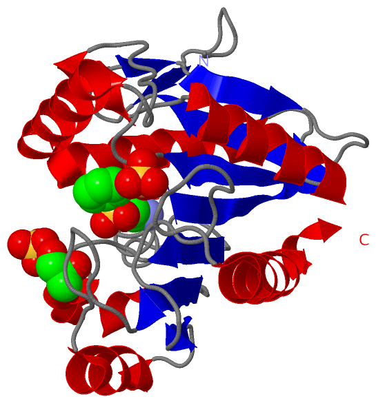
Ligands, Modified Residues, Ions (3, 4)| Asymmetric/Biological Unit (3, 4) |
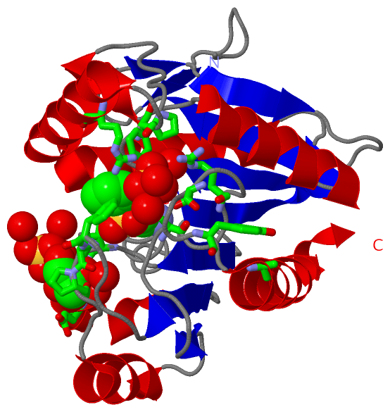
Sites (3, 3)
Asymmetric Unit (3, 3)
|
SS Bonds (0, 0)| (no "SS Bond" information available for 1ZJ5) |
Cis Peptide Bonds (1, 1)
Asymmetric/Biological Unit
|
||||||||
SAPs(SNPs)/Variants (0, 0)| (no "SAP(SNP)/Variant" information available for 1ZJ5) |
PROSITE Motifs (0, 0)| (no "PROSITE Motif" information available for 1ZJ5) |
Exons (0, 0)| (no "Exon" information available for 1ZJ5) |
Sequences/Alignments
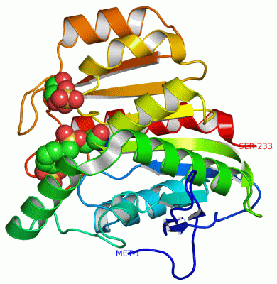
Asymmetric/Biological UnitChain A from PDB Type:PROTEIN Length:233 aligned with CLCD_PSEPU | P0A114 from UniProtKB/Swiss-Prot Length:236 Alignment length:233 10 20 30 40 50 60 70 80 90 100 110 120 130 140 150 160 170 180 190 200 210 220 230 CLCD_PSEPU 1 MLTEGISIQSYDGHTFGALVGSPAKAPAPVIVIAQEIFGVNAFMRETVSWLVDQGYAAVCPDLYARQAPGTALDPQDERQREQAYKLWQAFDMEAGVGDLEAAIRYARHQPYSNGKVGLVGYCLGGALAFLVAAKGYVDRAVGYYGVGLEKQLKKVPEVKHPALFHMGGQDHFVPAPSRQLITEGFGANPLLQVHWYEEAGHSFARTSSSGYVASAAALANERRLDFLAPLQS 233 SCOP domains d1zj5a_ A: automated matches SCOP domains CATH domains 1zj5A00 A:1-233 [code=3.40.50.1820, no name defined] CATH domains Pfam domains ---------------DLH-1zj5A01 A:16-232 - Pfam domains SAPs(SNPs) ----------------------------------------------------------------------------------------------------------------------------------------------------------------------------------------------------------------------------------------- SAPs(SNPs) PROSITE ----------------------------------------------------------------------------------------------------------------------------------------------------------------------------------------------------------------------------------------- PROSITE Transcript ----------------------------------------------------------------------------------------------------------------------------------------------------------------------------------------------------------------------------------------- Transcript 1zj5 A 1 MLTEGISIQSYDGHTFGALVGSPAKAPAPVIVIAQDIFGVNAFMRETVSWLVDQGYAAVCPDLYARQAPGTALDPQDERQREQAYKLWQAFDMEAGVGDLEAAIRYARHQPYSNGKVGLVGYsLGGALAFLVASKGYVDRAVGYYGVGLEKQLNKVPEVKHPALFHMGGQDHFVPAPSRQLITEGFGANPLLQVHWYEEAGHSFARTGSSGYVASAAALANERTLDFLVPLQS 233 10 20 30 40 50 60 70 80 90 100 110 120 | 130 140 150 160 170 180 190 200 210 220 230 123-SEB
|
||||||||||||||||||||
SCOP Domains (1, 1)
Asymmetric/Biological Unit
 |
CATH Domains (1, 1)
Asymmetric/Biological Unit
|
Pfam Domains (1, 1)| Asymmetric/Biological Unit |
Gene Ontology (4, 4)|
Asymmetric/Biological Unit(hide GO term definitions) Chain A (CLCD_PSEPU | P0A114)
|
||||||||||||||||||||||||||||||||||||
Interactive Views
|
|||||||||||||||||||||||||||||||||||||||||||||||||||||||||||||||||||||||||||||||||||||||||||||||||||||||||||||||||||||||||||||||||||||||||||||||||||
Still Images
|
||||||||||||||||
Databases
|
||||||||||||||||||||||||||||||||||||||||||||||||||||||||||||||||||||||||||||||||||||||||||||||||||||||||||||||||||||||||||||||||||||||||||||||||||||||||||||||||
Analysis Tools
|
|||||||||||||||||||||||||||||||||||||||||||||||||||||||||||||
Entries Sharing at Least One Protein Chain (UniProt ID)
Related Entries Specified in the PDB File
|
|