Show PDB file:   
         Plain Text   HTML   (compressed file size)
QuickSearch:   
by PDB,NDB,UniProt,PROSITE Code or Search Term(s)  
(-)Asymmetric Unit
(-)Asym. Unit - sites
(-)Biological Unit 1
(-)Biol. Unit 1 - sites
(-)Biological Unit 2
collapse expand < >
Image Asymmetric Unit
Asymmetric Unit  (Jmol Viewer)
Image Asym. Unit - sites
Asym. Unit - sites  (Jmol Viewer)
Image Biological Unit 1
Biological Unit 1  (Jmol Viewer)
Image Biol. Unit 1 - sites
Biol. Unit 1 - sites  (Jmol Viewer)
Image Biological Unit 2
Biological Unit 2  (Jmol Viewer)

(-) Description

Title :  CRYSTAL STRUCTURE OF EARTHWORM FIBRINOLYTIC ENZYME COMPONENT B: A NOVEL, GLYCOSYLATED TWO-CHAINED TRYPSIN
 
Authors :  F. Wang, C. Wang, M. Li, J. P. Zhang, L. L. Gui, X. M. An, W. R. Chang
Date :  20 Jan 05  (Deposition) - 19 Apr 05  (Release) - 13 Jul 11  (Revision)
Method :  X-RAY DIFFRACTION
Resolution :  2.06
Chains :  Asym. Unit :  A,B
Biol. Unit 1:  A,B  (1x)
Biol. Unit 2:  A,B  (2x)
Keywords :  Two Chains, Glycosylation, Pyroglutamation, Eight-Membered Ring, Cis Peptide Bond, Hydrolase (Keyword Search: [Gene Ontology, PubMed, Web (Google))
 
Reference :  F. Wang, C. Wang, M. Li, J. P. Zhang, L. L. Gui, X. M. An, W. R. Chang
Crystal Structure Of Earthworm Fibrinolytic Enzyme Componen B: A Novel, Glycosylated Two-Chained Trypsin.
J. Mol. Biol. V. 348 671 2005
PubMed-ID: 15826663  |  Reference-DOI: 10.1016/J.JMB.2005.02.055
(for further references see the PDB file header)

(-) Compounds

Molecule 1 - FIBRINOTIC ENZYME COMPONENT B
    ChainsA
    EC Number3.4.21.-
    FragmentRESIDUES 16-245
    Organism CommonCOMMON BRANDLING WORM
    Organism ScientificEISENIA FETIDA
    Organism Taxid6396
 
Molecule 2 - FIBRINOTIC ENZYME COMPONENT B
    ChainsB
    EC Number3.4.21.-
    FragmentRESIDUES 1J-13D
    Organism CommonCOMMON BRANDLING WORM
    Organism ScientificEISENIA FETIDA
    Organism Taxid6396

 Structural Features

(-) Chains, Units

  12
Asymmetric Unit AB
Biological Unit 1 (1x)AB
Biological Unit 2 (2x)AB

Summary Information (see also Sequences/Alignments below)

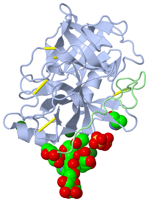
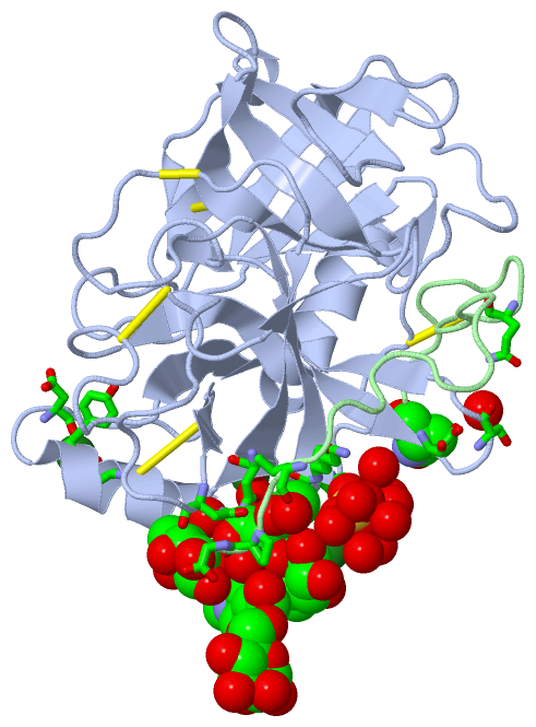
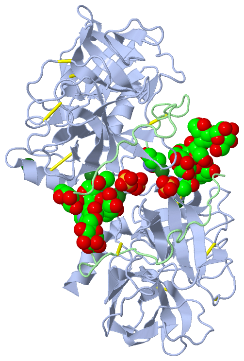
(-) Ligands, Modified Residues, Ions  (6, 12)

Asymmetric Unit (6, 12)
No.NameCountTypeFull Name
1FUC2Ligand/IonALPHA-L-FUCOSE
2MAN2Ligand/IonALPHA-D-MANNOSE
3MG3Ligand/IonMAGNESIUM ION
4NAG2Ligand/IonN-ACETYL-D-GLUCOSAMINE
5PCA1Mod. Amino AcidPYROGLUTAMIC ACID
6SO42Ligand/IonSULFATE ION
Biological Unit 1 (5, 9)
No.NameCountTypeFull Name
1FUC2Ligand/IonALPHA-L-FUCOSE
2MAN2Ligand/IonALPHA-D-MANNOSE
3MG-1Ligand/IonMAGNESIUM ION
4NAG2Ligand/IonN-ACETYL-D-GLUCOSAMINE
5PCA1Mod. Amino AcidPYROGLUTAMIC ACID
6SO42Ligand/IonSULFATE ION
Biological Unit 2 (5, 18)
No.NameCountTypeFull Name
1FUC4Ligand/IonALPHA-L-FUCOSE
2MAN4Ligand/IonALPHA-D-MANNOSE
3MG-1Ligand/IonMAGNESIUM ION
4NAG4Ligand/IonN-ACETYL-D-GLUCOSAMINE
5PCA2Mod. Amino AcidPYROGLUTAMIC ACID
6SO44Ligand/IonSULFATE ION

(-) Sites  (11, 11)

Asymmetric Unit (11, 11)
No.NameEvidenceResiduesDescription
01AC1SOFTWAREFUC A:2 , FUC A:3 , NAG A:4 , TYR A:133 , LYS A:135 , ASN A:161 , ASN A:186 , HOH A:559 , LEU B:13A , HOH B:18BINDING SITE FOR RESIDUE NAG A 1
02AC2SOFTWARENAG A:1 , FUC A:2 , MAN A:5 , HOH A:571 , PRO B:13C , HOH B:34BINDING SITE FOR RESIDUE NAG A 4
03AC3SOFTWARENAG A:4 , MAN A:6 , GLN B:1ABINDING SITE FOR RESIDUE MAN A 5
04AC4SOFTWARENAG A:1 , NAG A:4 , ASP A:203 , SER A:204A , HOH A:505 , HOH A:506 , HOH A:574 , HOH A:640BINDING SITE FOR RESIDUE FUC A 2
05AC5SOFTWARENAG A:1 , ASN A:186 , HOH A:653 , PRO B:13C , GLY B:13DBINDING SITE FOR RESIDUE FUC A 3
06AC6SOFTWAREMAN A:5BINDING SITE FOR RESIDUE MAN A 6
07AC7SOFTWAREHOH A:503 , HOH A:504 , HOH A:505 , HOH A:506 , HOH A:507 , HOH A:508BINDING SITE FOR RESIDUE MG A 401
08AC8SOFTWAREASP A:169 , TYR A:172 , ASP A:174ABINDING SITE FOR RESIDUE MG A 402
09AC9SOFTWAREHOH A:572 , HOH A:599 , HOH A:624 , HOH A:625 , HOH A:626 , HOH A:627BINDING SITE FOR RESIDUE MG A 403
10BC1SOFTWAREARG A:134 , LYS A:135 , HOH A:503 , HOH A:507 , HOH A:550 , HOH A:574 , HOH A:602 , HOH A:603 , HOH A:677 , HOH A:684BINDING SITE FOR RESIDUE SO4 A 501
11BC2SOFTWAREASN A:128 , ARG A:134 , HOH A:564BINDING SITE FOR RESIDUE SO4 A 502

(-) SS Bonds  (5, 5)

Asymmetric Unit
No.Residues
1A:42 -A:58
2A:122 -B:1
3A:150 -A:151
4A:168 -A:182
5A:191 -A:220

(-) Cis Peptide Bonds  (1, 1)

Asymmetric Unit
No.Residues
1Cys A:150 -Cys A:151

 Sequence-Structure Mapping

(-) SAPs(SNPs)/Variants  (0, 0)

(no "SAP(SNP)/Variant" information available for 1YM0)

(-) PROSITE Motifs  (0, 0)

(no "PROSITE Motif" information available for 1YM0)

(-) Exons   (0, 0)

(no "Exon" information available for 1YM0)

(-) Sequences/Alignments

Asymmetric Unit
   Reformat: Number of residues per line =  ('0' or empty: single-line sequence representation)
  Number of residues per labelling interval =   
  UniProt sequence: complete  aligned part    
   Show mapping: SCOP domains CATH domains Pfam domains Secondary structure (by author)
SAPs(SNPs) PROSITE motifs Exons
(details for a mapped element are shown in a popup box when the mouse pointer rests over it)
Chain A from PDB  Type:PROTEIN  Length:238
 aligned with Q3HR18_EISFE | Q3HR18 from UniProtKB/TrEMBL  Length:245

    Alignment length:238
                                    17        27        37        47        57        67        77        87        97       107       117       127       137       147       157       167       177       187       197       207       217       227       237        
        Q3HR18_EISFE      8 IVGGIEARPYEFPWQVSVRRKSSDSHFCGGSIINDRWVACAAHCMQGESPALVSLVVGEHDSSAASTVRQTHDVDSIFVNENYDPRTLENDVSVIKTAIAITFDINVGPICAPDPANDYVYRKSQCSGWGTINSGGICCPAVLRYVTLNITTNAFCDAVYTSDTIYDDMICATDNTGMTDRDSCQGDSGGPLSVKDGSGIFSLVGIVSWGIGCASGYPGVYSRVGFHAGWITDTITNN  245
               SCOP domains d1ym0a_ A: automated matches                                                                                                                                                                                                                   SCOP domains
               CATH domains 1ym0A01     1ym0A02 A:28-120,A:231-245 Trypsin-like serine proteases                                         1ym0A01 A:16-27,A:121-230 Trypsin-like serine proteases                                                           1ym0A02         CATH domains
               Pfam domains ---------------------------------------------------------------------------------------------------------------------------------------------------------------------------------------------------------------------------------------------- Pfam domains
         Sec.struct. author ....ee........eeeeee....eeeeeeeeee..eeeehhhhhh..hhh.eeeee............eeeeeeeeee.............eeeee.........................eeeeee...............eeeeeee.hhhhhhhhh........eeeee.................eeeee.....eeeeeeeee.........eeeeehhhhhhhhhhhhhh. Sec.struct. author
                 SAPs(SNPs) ---------------------------------------------------------------------------------------------------------------------------------------------------------------------------------------------------------------------------------------------- SAPs(SNPs)
                    PROSITE ---------------------------------------------------------------------------------------------------------------------------------------------------------------------------------------------------------------------------------------------- PROSITE
                 Transcript ---------------------------------------------------------------------------------------------------------------------------------------------------------------------------------------------------------------------------------------------- Transcript
                1ym0 A   16 IVGGIEARPYEFPWQVSVRRKSSDSHFCGGSIINDRWVVCAAHCMQGEAPALVSLVVGEHDSSAASTVRQTHDVDSIFVNENYDPATLENDVSVIKTAVAITFDINVGPICAPDPANDYVYRKSQCSGWGTINSGGVCCPAVLRYVTLNITTNAFCDAVYTSDTIYDDMICATDNTGMTDRDSCQGDSGGPLSVKDGSGIFSLVGIVSWGIGCASGYPGVYSRVGFHAGWITDTITNN  245
                                    25        35  |     44        54      ||61        71        81        91       101       111       121       131|      142       152       162       172  |    181     ||187       197       206       216||     227       237        
                                                37A                     60A||                                                                    131|                                      174A         186A|||               204A          217|                          
                                                                         60B|                                                                     133                                                    186B||                              219                          
                                                                          60C                                                                                                                             186C|                                                           
                                                                                                                                                                                                           186D                                                           

Chain B from PDB  Type:PROTEIN  Length:27
                                                            
               SCOP domains --------------------------- SCOP domains
               CATH domains --------------------------- CATH domains
               Pfam domains --------------------------- Pfam domains
         Sec.struct. author ........................... Sec.struct. author
                 SAPs(SNPs) --------------------------- SAPs(SNPs)
                    PROSITE --------------------------- PROSITE
                 Transcript --------------------------- Transcript
                1ym0 B   1J xPPVWYPGGQCGVSQYSDAGDMELPPG  13D
                            ||||||||1A        10   ||||
                           1J-PCA|||1A           13A|||
                            1I|||||||             13B||
                             1H||||||              13C|
                              1G|||||               13D
                               1F||||                  
                                1E|||                  
                                 1D||                  
                                  1C|                  
                                   1B                  

   Legend:   → Mismatch (orange background)
  - → Gap (green background, '-', border residues have a numbering label)
    → Modified Residue (blue background, lower-case, 'x' indicates undefined single-letter code, labelled with number + name)
  x → Chemical Group (purple background, 'x', labelled with number + name, e.g. ACE or NH2)
  extra numbering lines below/above indicate numbering irregularities and modified residue names etc., number ends below/above '|'

 Classification and Annotation

(-) SCOP Domains  (1, 1)

Asymmetric Unit

(-) CATH Domains  (1, 2)

Asymmetric Unit
(-)
Class: Mainly Beta (13760)

(-) Pfam Domains  (0, 0)

(no "Pfam Domain" information available for 1YM0)

(-) Gene Ontology  (5, 5)

Asymmetric Unit(hide GO term definitions)
Chain A   (Q3HR18_EISFE | Q3HR18)
molecular function
    GO:0016301    kinase activity    Catalysis of the transfer of a phosphate group, usually from ATP, to a substrate molecule.
    GO:0046872    metal ion binding    Interacting selectively and non-covalently with any metal ion.
    GO:0004252    serine-type endopeptidase activity    Catalysis of the hydrolysis of internal, alpha-peptide bonds in a polypeptide chain by a catalytic mechanism that involves a catalytic triad consisting of a serine nucleophile that is activated by a proton relay involving an acidic residue (e.g. aspartate or glutamate) and a basic residue (usually histidine).
biological process
    GO:0016310    phosphorylation    The process of introducing a phosphate group into a molecule, usually with the formation of a phosphoric ester, a phosphoric anhydride or a phosphoric amide.
    GO:0006508    proteolysis    The hydrolysis of proteins into smaller polypeptides and/or amino acids by cleavage of their peptide bonds.

 Visualization

(-) Interactive Views

Asymmetric Unit
  Complete Structure
    Jena3D(integrated viewing of ligand, site, SAP, PROSITE, SCOP information)
    WebMol | AstexViewer[tm]@PDBe
(Java Applets, require no local installation except for Java; loading may be slow)
    STRAP
(Java WebStart application, automatic local installation, requires Java; full application with system access!)
    RasMol
(require local installation)
    Molscript (VRML)
(requires installation of a VRML viewer; select preferred view via VRML and generate a mono or stereo PDF format file)
 
  Ligands, Modified Residues, Ions
    FUC  [ RasMol | Jena3D ]  +environment [ RasMol | Jena3D ]
    MAN  [ RasMol | Jena3D ]  +environment [ RasMol | Jena3D ]
    MG  [ RasMol | Jena3D ]  +environment [ RasMol | Jena3D ]
    NAG  [ RasMol | Jena3D ]  +environment [ RasMol | Jena3D ]
    PCA  [ RasMol | Jena3D ]  +environment [ RasMol | Jena3D ]
    SO4  [ RasMol | Jena3D ]  +environment [ RasMol | Jena3D ]
 
  Sites
    AC1  [ RasMol ]  +environment [ RasMol ]
    AC2  [ RasMol ]  +environment [ RasMol ]
    AC3  [ RasMol ]  +environment [ RasMol ]
    AC4  [ RasMol ]  +environment [ RasMol ]
    AC5  [ RasMol ]  +environment [ RasMol ]
    AC6  [ RasMol ]  +environment [ RasMol ]
    AC7  [ RasMol ]  +environment [ RasMol ]
    AC8  [ RasMol ]  +environment [ RasMol ]
    AC9  [ RasMol ]  +environment [ RasMol ]
    BC1  [ RasMol ]  +environment [ RasMol ]
    BC2  [ RasMol ]  +environment [ RasMol ]
 
  Cis Peptide Bonds
    Cys A:150 - Cys A:151   [ RasMol ]  
 
Biological Units
  Complete Structure
    Biological Unit 1  [ Jena3D ]
    Biological Unit 2  [ Jena3D ]

(-) Still Images

Jmol
  protein: cartoon or spacefill or dots and stick; nucleic acid: cartoon and stick; ligands: spacefill; active site: stick
Molscript
  protein, nucleic acid: cartoon; ligands: spacefill; active site: ball and stick

 Databases and Analysis Tools

(-) Databases

Access by PDB/NDB ID
  1ym0
    Family and Domain InformationProDom | SYSTERS
    General Structural InformationGlycoscienceDB | MMDB | NDB | OCA | PDB | PDBe | PDBj | PDBsum | PDBWiki | PQS | PROTEOPEDIA
    Orientation in MembranesOPM
    Protein SurfaceSURFACE
    Secondary StructureDSSP (structure derived) | HSSP (homology derived)
    Structural GenomicsGeneCensus
    Structural NeighboursCE | VAST
    Structure ClassificationCATH | Dali | SCOP
    Validation and Original DataBMRB Data View | BMRB Restraints Grid | EDS | PROCHECK | RECOORD | WHAT_CHECK
 
Access by UniProt ID/Accession number
  Q3HR18_EISFE | Q3HR18
    Comparative Protein Structure ModelsModBase
    Genomic InformationEnsembl
    Protein-protein InteractionDIP
    Sequence, Family and Domain InformationInterPro | Pfam | SMART | UniProtKB/TrEMBL
 
Access by Enzyme Classificator   (EC Number)
  3.4.21.-
    General Enzyme InformationBRENDA | EC-PDB | Enzyme | IntEnz
    PathwayKEGG | MetaCyc
 
Access by Disease Identifier   (MIM ID)
  (no 'MIM ID' available)
    Disease InformationOMIM
 
Access by GenAge ID
  (no 'GenAge ID' available)
    Age Related InformationGenAge

(-) Analysis Tools

Access by PDB/NDB ID
    Domain InformationXDom
    Interatomic Contacts of Structural UnitsCSU
    Ligand-protein ContactsLPC
    Protein CavitiescastP
    Sequence and Secondary StructurePDBCartoon
    Structure AlignmentSTRAP(Java WebStart application, automatic local installation, requires Java; full application with system access!)
    Structure and Sequence BrowserSTING
 
Access by UniProt ID/Accession number
  Q3HR18_EISFE | Q3HR18
    Protein Disorder PredictionDisEMBL | FoldIndex | GLOBPLOT (for more information see DisProt)

 Related Entries

(-) Entries Sharing at Least One Protein Chain (UniProt ID)

(no "Entries Sharing at Least One Protein Chain" available for 1YM0)

(-) Related Entries Specified in the PDB File

(no "Related Entries Specified in the PDB File" available for 1YM0)