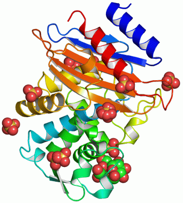
Asymmetric Unit (11, 11)
| No. | Name | Evidence | Residues | Description |
|---|
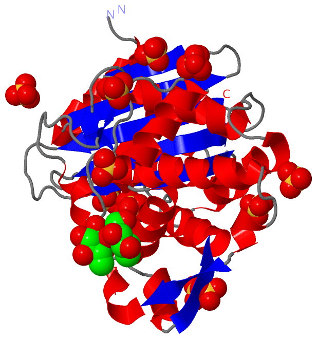
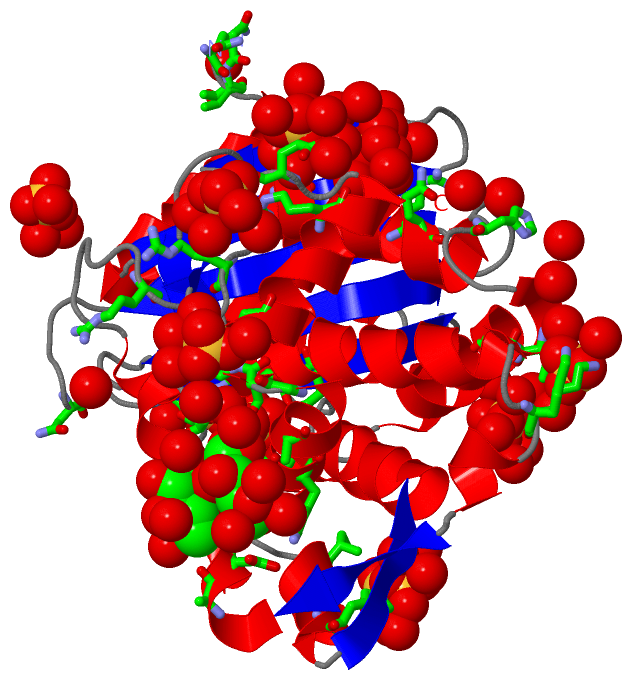
| 01 | AC1 | SOFTWARE | ALA A:100 , ASP A:101 , ASN A:136 , LYS A:137 , ALA A:140 , THR A:165 , SO4 A:1008 , HOH A:2001 , HOH A:2044 , HOH A:2088 , HOH A:2212 , HOH A:2363 , HOH A:2394 , HOH A:2428 , HOH A:2448 , HOH A:2473 | BINDING SITE FOR RESIDUE SUC A 1050 |
| 02 | AC2 | SOFTWARE | ARG A:153 , ARG A:191 , HIS A:197 , HOH A:2081 , HOH A:2103 , HOH A:2151 , HOH A:2342 , HOH A:2365 , HOH A:2421 | BINDING SITE FOR RESIDUE SO4 A 1001 |
| 03 | AC3 | SOFTWARE | SER A:70 , SER A:130 , LYS A:234 , THR A:235 , GLY A:236 , SER A:237 , HOH A:2017 , HOH A:2018 , HOH A:2122 , HOH A:2226 , HOH A:2303 , HOH A:2438 | BINDING SITE FOR RESIDUE SO4 A 1002 |
| 04 | AC4 | SOFTWARE | ARG A:161 , GLN A:188 , GLN A:192 , HOH A:2037 , HOH A:2076 , HOH A:2138 , HOH A:2155 , HOH A:2210 , HOH A:2225 , HOH A:2324 , HOH A:2375 | BINDING SITE FOR RESIDUE SO4 A 1003 |
| 05 | AC5 | SOFTWARE | ARG A:184 , GLN A:188 , HOH A:2012 , HOH A:2106 , HOH A:2180 , HOH A:2219 , HOH A:2225 , HOH A:2248 , HOH A:2374 , HOH A:2432 | BINDING SITE FOR RESIDUE SO4 A 1004 |
| 06 | AC6 | SOFTWARE | LYS A:88 , ARG A:178 , THR A:202 , GLN A:203 , HOH A:2063 , HOH A:2128 , HOH A:2397 , HOH A:2479 | BINDING SITE FOR RESIDUE SO4 A 1005 |
| 07 | AC7 | SOFTWARE | GLY A:143 , GLY A:144 , PRO A:145 , GLY A:146 , HOH A:2296 , HOH A:2402 , HOH A:2419 , HOH A:2466 , HOH A:2475 | BINDING SITE FOR RESIDUE SO4 A 1006 |
| 08 | AC8 | SOFTWARE | SER A:274 , ARG A:276 , ASP A:277 , HOH A:2055 , HOH A:2200 , HOH A:2344 | BINDING SITE FOR RESIDUE SO4 A 1007 |
| 09 | AC9 | SOFTWARE | ILE A:108 , LYS A:111 , HIS A:112 , GLU A:288 , SUC A:1050 , HOH A:2056 , HOH A:2071 , HOH A:2119 , HOH A:2240 , HOH A:2270 , HOH A:2377 , HOH A:2473 | BINDING SITE FOR RESIDUE SO4 A 1008 |
| 10 | BC1 | SOFTWARE | GLN A:25 , THR A:26 , ASN A:271 , HOH A:2084 , HOH A:2113 , HOH A:2326 , HOH A:2336 , HOH A:2339 , HOH A:2371 , HOH A:2381 | BINDING SITE FOR RESIDUE SO4 A 1009 |
| 11 | BC2 | SOFTWARE | ARG A:153 , GLU A:158 , HIS A:197 , HOH A:2080 , HOH A:2091 , HOH A:2227 , HOH A:2337 , HOH A:2342 | BINDING SITE FOR RESIDUE SO4 A 1010 |
|
 Description