|
|
|
|
 Description
|
|
Compounds
|
||||||||||||||||||||||||||||||||||||||||||||||||||||
Chains, Units
Summary Information (see also Sequences/Alignments below) |
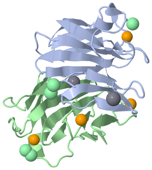
Ligands, Modified Residues, Ions (3, 10)| Asymmetric/Biological Unit (3, 10) |
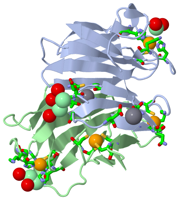
Sites (10, 10)
Asymmetric Unit (10, 10)
|
SS Bonds (0, 0)| (no "SS Bond" information available for 1VZI) |
Cis Peptide Bonds (2, 2)
Asymmetric/Biological Unit
|
||||||||||||
SAPs(SNPs)/Variants (0, 0)| (no "SAP(SNP)/Variant" information available for 1VZI) |
PROSITE Motifs (0, 0)| (no "PROSITE Motif" information available for 1VZI) |
Exons (0, 0)| (no "Exon" information available for 1VZI) |
Sequences/Alignments
Asymmetric/Biological UnitChain A from PDB Type:PROTEIN Length:125 aligned with DFX_DESB2 | Q46495 from UniProtKB/Swiss-Prot Length:126 Alignment length:125 11 21 31 41 51 61 71 81 91 101 111 121 DFX_DESB2 2 PERLQVYKCEVCGNIVEVLNGGIGELVCCNQDMKLMSENTVDAAKEKHVPVIEKIDGGYKVKVGAVAHPMEEKHYIQWIELLADDKCYTQFLKPGQAPEAVFLIEAAKVVAREYCNIHGHWKAEN 126 SCOP domains d1vzia2 A:1-37 d1vzia1 A:38-125 Desulfoferrodoxin C-terminal domain SCOP domains CATH domains ----------------------------------------------------------------------------------------------------------------------------- CATH domains Pfam domains ----------------------------------------------------------------------------------------------------------------------------- Pfam domains SAPs(SNPs) ----------------------------------------------------------------------------------------------------------------------------- SAPs(SNPs) PROSITE ----------------------------------------------------------------------------------------------------------------------------- PROSITE Transcript ----------------------------------------------------------------------------------------------------------------------------- Transcript 1vzi A 1 PERLQVYKCEVCGNIVEVLNGGIGELVCCNQDMKLMSENTVDAAKAKHVPVIEKIDGGYKVKVGAVAHPMEEKHYIQWIELLADDKCYTQFLKPGQAPEAVFLIEAAKVVAREYCNIHGHWKAEN 125 10 20 30 40 50 60 70 80 90 100 110 120 Chain B from PDB Type:PROTEIN Length:125 aligned with DFX_DESB2 | Q46495 from UniProtKB/Swiss-Prot Length:126 Alignment length:125 11 21 31 41 51 61 71 81 91 101 111 121 DFX_DESB2 2 PERLQVYKCEVCGNIVEVLNGGIGELVCCNQDMKLMSENTVDAAKEKHVPVIEKIDGGYKVKVGAVAHPMEEKHYIQWIELLADDKCYTQFLKPGQAPEAVFLIEAAKVVAREYCNIHGHWKAEN 126 SCOP domains d1vzib2 B:1-37 d1vzib1 B:38-125 Desulfoferrodoxin C-terminal domain SCOP domains CATH domains ----------------------------------------------------------------------------------------------------------------------------- CATH domains Pfam domains (1) Desulfoferrod_N-1vziB01 B:1-36 ---Desulfoferrodox-1vziB03 B:40-124 - Pfam domains (1) Pfam domains (2) Desulfoferrod_N-1vziB02 B:1-36 ---Desulfoferrodox-1vziB04 B:40-124 - Pfam domains (2) SAPs(SNPs) ----------------------------------------------------------------------------------------------------------------------------- SAPs(SNPs) PROSITE ----------------------------------------------------------------------------------------------------------------------------- PROSITE Transcript ----------------------------------------------------------------------------------------------------------------------------- Transcript 1vzi B 1 PERLQVYKCEVCGNIVEVLNGGIGELVCCNQDMKLMSENTVDAAKAKHVPVIEKIDGGYKVKVGAVAHPMEEKHYIQWIELLADDKCYTQFLKPGQAPEAVFLIEAAKVVAREYCNIHGHWKAEN 125 10 20 30 40 50 60 70 80 90 100 110 120
|
||||||||||||||||||||
SCOP Domains (2, 4)
Asymmetric/Biological Unit
 |
CATH Domains (0, 0)| (no "CATH Domain" information available for 1VZI) |
Pfam Domains (2, 4)| Asymmetric/Biological Unit |
Gene Ontology (7, 7)|
Asymmetric/Biological Unit(hide GO term definitions) Chain A,B (DFX_DESB2 | Q46495)
|
||||||||||||||||||||||||||||||||||||||||||||||||||||||
Interactive Views
|
|||||||||||||||||||||||||||||||||||||||||||||||||||||||||||||||||||||||||||||||||||||||||||||||||||||||||||||||||||||||||||||||||||||||||||||||||||||||||||||||||||||||||||||||||||||||||||||||||||||||||||
Still Images
|
||||||||||||||||
Databases
|
||||||||||||||||||||||||||||||||||||||||||||||||||||||||||||||||||||||||||||||||||||||||||||||||||||||||||||||||||||||||||||||||||||||||||||||||||||||||||||||||
Analysis Tools
|
|||||||||||||||||||||||||||||||||||||||||||||||||||||||||||||
Entries Sharing at Least One Protein Chain (UniProt ID)
Related Entries Specified in the PDB File
|
|