Show PDB file:   
         Plain Text   HTML   (compressed file size)
QuickSearch:   
by PDB,NDB,UniProt,PROSITE Code or Search Term(s)  
(-)Asym./Biol. Unit
(-)Asym./Biol. Unit - sites
collapse expand < >
Image Asym./Biol. Unit
Asym./Biol. Unit  (Jmol Viewer)
Image Asym./Biol. Unit - sites
Asym./Biol. Unit - sites  (Jmol Viewer)

(-) Description

Title :  A SECOND LOOK AT THE HDV RIBOZYME STRUCTURE AND DYNAMICS.
 
Authors :  G. J. Kapral, S. Jain, J. Noeske, J. A. Doudna, D. C. Richardson, J. S. Ric
Date :  05 Mar 14  (Deposition) - 29 Oct 14  (Release) - 26 Nov 14  (Revision)
Method :  X-RAY DIFFRACTION
Resolution :  2.40
Chains :  Asym./Biol. Unit :  A,B
Keywords :  Hdv, Ribozyme, Rna, U1A, Precursor, Translation-Rna Complex, Rna Binding Protein-Rna Complex (Keyword Search: [Gene Ontology, PubMed, Web (Google))
 
Reference :  G. J. Kapral, S. Jain, J. Noeske, J. A. Doudna, D. C. Richardson, J. S. Richardson
New Tools Provide A Second Look At Hdv Ribozyme Structure, Dynamics And Cleavage.
Nucleic Acids Res. V. 42 12833 2014
PubMed-ID: 25326328  |  Reference-DOI: 10.1093/NAR/GKU992

(-) Compounds

Molecule 1 - HEPATITIS DELTA VIRUS RIBOZYME
    ChainsB
    EngineeredYES
    Other DetailsRNA OCCURS FROM HAPATITIS DELTA VIRUS PATHOGEN, IN VITRO TRANSCRIPTION WITH PUC19
    SyntheticYES
 
Molecule 2 - U1 SMALL NUCLEAR RIBONUCLEOPROTEIN A
    ChainsA
    EngineeredYES
    Expression SystemESCHERICHIA COLI
    Expression System PlasmidPET
    Expression System Taxid562
    Expression System Vector TypePLASMID
    FragmentU1A_RBD, UNP RESIDUES 98-173
    GeneSNRPA
    MutationYES
    Organism CommonHUMAN
    Organism ScientificHOMO SAPIENS
    Organism Taxid9606
    SynonymU1 SNRNP A, U1-A, U1A

 Structural Features

(-) Chains, Units

  12
Asymmetric/Biological Unit AB

Summary Information (see also Sequences/Alignments below)

(-) Ligands, Modified Residues, Ions  (1, 2)

Asymmetric/Biological Unit (1, 2)
No.NameCountTypeFull Name
1SR2Ligand/IonSTRONTIUM ION

(-) Sites  (2, 2)

Asymmetric Unit (2, 2)
No.NameEvidenceResiduesDescription
1AC1SOFTWAREU B:120 , U B:123 , U B:163BINDING SITE FOR RESIDUE SR B 201
2AC2SOFTWAREC B:141BINDING SITE FOR RESIDUE SR B 202

(-) SS Bonds  (0, 0)

(no "SS Bond" information available for 4PRF)

(-) Cis Peptide Bonds  (0, 0)

(no "Cis Peptide Bond" information available for 4PRF)

 Sequence-Structure Mapping

(-) SAPs(SNPs)/Variants  (0, 0)

(no "SAP(SNP)/Variant" information available for 4PRF)

(-) PROSITE Motifs  (0, 0)

(no "PROSITE Motif" information available for 4PRF)

(-) Exons   (0, 0)

(no "Exon" information available for 4PRF)

(-) Sequences/Alignments

Asymmetric/Biological Unit
   Reformat: Number of residues per line =  ('0' or empty: single-line sequence representation)
  Number of residues per labelling interval =   
  UniProt sequence: complete  aligned part    
   Show mapping: SCOP domains CATH domains Pfam domains Secondary structure (by author)
SAPs(SNPs) PROSITE motifs Exons
(details for a mapped element are shown in a popup box when the mouse pointer rests over it)
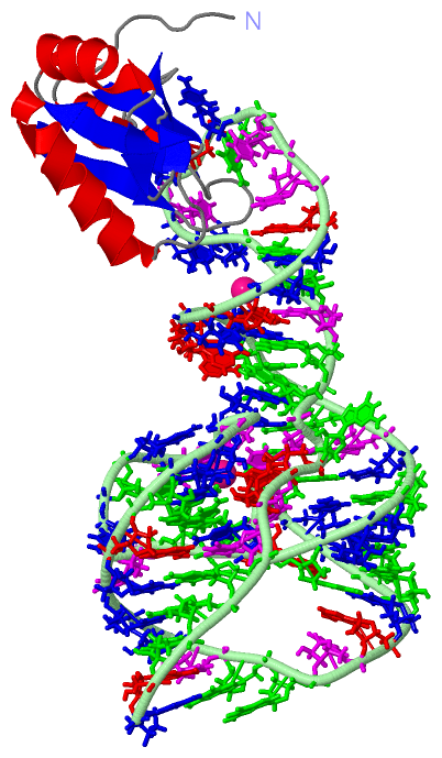
Chain A from PDB  Type:PROTEIN  Length:95
                                                                                                                               
               SCOP domains ----------------------------------------------------------------------------------------------- SCOP domains
               CATH domains ----------------------------------------------------------------------------------------------- CATH domains
               Pfam domains ----------------------------------------------------------------------------------------------- Pfam domains
         Sec.struct. author .......eeeee......hhhhhhhhhhhhhhhhh.eeeeee.........eeeee.hhhhhhhhhhhhh..ee..ee.eeee....hhhhhh.. Sec.struct. author
                 SAPs(SNPs) ----------------------------------------------------------------------------------------------- SAPs(SNPs)
                    PROSITE ----------------------------------------------------------------------------------------------- PROSITE
                 Transcript ----------------------------------------------------------------------------------------------- Transcript
                 4prf A   4 PETRPNHTIYINNLNEKIKKDELKKSLHAIFSRFGQILDILVSRSLKMRGQAFVIFKEVSSATNALRSMQGFPFYDKPMRIQYAKTDSDIIAKMK  98
                                    13        23        33        43        53        63        73        83        93     

Chain B from PDB  Type:RNA  Length:74
                                                                                                          
                 4prf B  99 AUGGCCGGCAUGGUCCCAGCCUCCUCGCUGGCGCCGGCUGGGCAACACCAUUGCACUCCGGUGGUGAAUGGGAC 172
                                   108       118       128       138       148       158       168    

   Legend:   → Mismatch (orange background)
  - → Gap (green background, '-', border residues have a numbering label)
    → Modified Residue (blue background, lower-case, 'x' indicates undefined single-letter code, labelled with number + name)
  x → Chemical Group (purple background, 'x', labelled with number + name, e.g. ACE or NH2)
  extra numbering lines below/above indicate numbering irregularities and modified residue names etc., number ends below/above '|'

 Classification and Annotation

(-) SCOP Domains  (0, 0 ; only for superseded entry 1VC7: 1,1)

(no "SCOP Domain" information available for 4PRF, only for superseded entry 1VC7 replaced by 4PRF)

(-) CATH Domains  (0, 0 ; only for superseded entry 1VC7: 1,1)

(no "CATH Domain" information available for 4PRF, only for superseded entry 1VC7 replaced by 4PRF)

(-) Pfam Domains  (0, 0)

(no "Pfam Domain" information available for 4PRF)

(-) Gene Ontology  (16, 16)

Asymmetric/Biological Unit(hide GO term definitions)

 Visualization

(-) Interactive Views

Asymmetric/Biological Unit
  Complete Structure
    Jena3D(integrated viewing of ligand, site, SAP, PROSITE, SCOP information)
    WebMol | AstexViewer[tm]@PDBe
(Java Applets, require no local installation except for Java; loading may be slow)
    STRAP
(Java WebStart application, automatic local installation, requires Java; full application with system access!)
    RasMol
(require local installation)
    Molscript (VRML)
(requires installation of a VRML viewer; select preferred view via VRML and generate a mono or stereo PDF format file)
 
  Ligands, Modified Residues, Ions
    SR  [ RasMol | Jena3D ]  +environment [ RasMol | Jena3D ]
 
  Sites
    AC1  [ RasMol ]  +environment [ RasMol ]
    AC2  [ RasMol ]  +environment [ RasMol ]
 
  Cis Peptide Bonds
(no "Cis Peptide Bonds" information available for 4prf)
 

(-) Still Images

Jmol
  protein: cartoon or spacefill or dots and stick; nucleic acid: cartoon and stick; ligands: spacefill; active site: stick
Molscript
  protein, nucleic acid: cartoon; ligands: spacefill; active site: ball and stick

 Databases and Analysis Tools

(-) Databases

Access by PDB/NDB ID
  4prf
    Family and Domain InformationProDom | SYSTERS
    General Structural InformationGlycoscienceDB | MMDB | NDB | OCA | PDB | PDBe | PDBj | PDBsum | PDBWiki | PQS | PROTEOPEDIA
    Orientation in MembranesOPM
    Protein SurfaceSURFACE
    Secondary StructureDSSP (structure derived) | HSSP (homology derived)
    Structural GenomicsGeneCensus
    Structural NeighboursCE | VAST
    Structure ClassificationCATH | Dali | SCOP
    Validation and Original DataBMRB Data View | BMRB Restraints Grid | EDS | PROCHECK | RECOORD | WHAT_CHECK
 
Access by UniProt ID/Accession number
  SNRPA_HUMAN | P09012
    Comparative Protein Structure ModelsModBase
    Genomic InformationEnsembl
    Protein-protein InteractionDIP
    Sequence, Family and Domain InformationInterPro | Pfam | SMART | UniProtKB/SwissProt
 
Access by Enzyme Classificator   (EC Number)
  (no 'Enzyme Classificator' available)
    General Enzyme InformationBRENDA | EC-PDB | Enzyme | IntEnz
    PathwayKEGG | MetaCyc
 
Access by Disease Identifier   (MIM ID)
  (no 'MIM ID' available)
    Disease InformationOMIM
 
Access by GenAge ID
  (no 'GenAge ID' available)
    Age Related InformationGenAge

(-) Analysis Tools

Access by PDB/NDB ID
    Domain InformationXDom
    Interatomic Contacts of Structural UnitsCSU
    Ligand-protein ContactsLPC
    Protein CavitiescastP
    Sequence and Secondary StructurePDBCartoon
    Structure AlignmentSTRAP(Java WebStart application, automatic local installation, requires Java; full application with system access!)
    Structure and Sequence BrowserSTING
 
Access by UniProt ID/Accession number
  SNRPA_HUMAN | P09012
    Protein Disorder PredictionDisEMBL | FoldIndex | GLOBPLOT (for more information see DisProt)

 Related Entries

(-) Entries Sharing at Least One Protein Chain (UniProt ID)

UniProtKB/Swiss-Prot
        SNRPA_HUMAN | P090121aud 1drz 1dz5 1fht 1m5k 1m5o 1m5p 1m5v 1nu4 1oia 1sj3 1sj4 1sjf 1u6b 1urn 1vbx 1vby 1vbz 1vc0 1vc5 1vc6 1zzn 2a3j 2nz4 2oih 2oj3 2u1a 3bo2 3bo3 3bo4 3cul 3cun 3egz 3g8s 3g8t 3g96 3g9c 3hhn 3iin 3irw 3iwn 3k0j 3l3c 3mum 3mur 3mut 3muv 3mxh 3p49 3pgw 3r1h 3r1l 3ucu 3ucz 3ud3 3ud4 3utr 4c4w 4pr6 4w90 4w92 4yb1 5ddo 5ddp 5ddq 5ddr 5fj4

(-) Related Entries Specified in the PDB File

4pr6 THE POST-CLEAVED HDV RIBOZYME REREFINED WITH ERRASER AND PHENIX