Show PDB file:   
         Plain Text   HTML   (compressed file size)
QuickSearch:   
by PDB,NDB,UniProt,PROSITE Code or Search Term(s)  
(-)Asym./Biol. Unit - manually
(-)Asym./Biol. Unit
(-)Asym./Biol. Unit - sites
collapse expand < >
Image Asym./Biol. Unit - manually
Asym./Biol. Unit - manually  (Jmol Viewer)
Image Asym./Biol. Unit
Asym./Biol. Unit  (Jmol Viewer)
Image Asym./Biol. Unit - sites
Asym./Biol. Unit - sites  (Jmol Viewer)

(-) Description

Title :  CRYSTAL STRUCTURE OF THE BROADLY NEUTRALIZING ANTI-HIV-1 ANTIBODY 2F5 IN COMPLEX WITH A GP41 7MER EPITOPE
 
Authors :  G. Ofek, M. Tang, A. Sambor, H. Katinger, J. R. Mascola, R. Wyatt, P. D. Kwong
Date :  04 Jun 04  (Deposition) - 05 Oct 04  (Release) - 24 Feb 09  (Revision)
Method :  X-RAY DIFFRACTION
Resolution :  2.00
Chains :  Asym./Biol. Unit :  H,L,P
Keywords :  2F5; Antibody; Gp41; Hiv-1; Neutralizing; Membrane-Proximal, Viral Protein/Immune System Complex (Keyword Search: [Gene Ontology, PubMed, Web (Google))
 
Reference :  G. Ofek, M. Tang, A. Sambor, H. Katinger, J. R. Mascola, R. Wyatt, P. D. Kwong
Structure And Mechanistic Analysis Of The Anti-Human Immunodeficiency Virus Type 1 Antibody 2F5 In Complex With Its Gp41 Epitope
J. Virol. V. 78 10724 2004
PubMed-ID: 15367639  |  Reference-DOI: 10.1128/JVI.78.19.10724-10737.2004
(for further references see the PDB file header)

(-) Compounds

Molecule 1 - FAB 2F5 LIGHT CHAIN
    ChainsL
    EngineeredYES
    Expression SystemCRICETULUS GRISEUS
    Expression System Cell LineCB-F7
    Expression System CommonCHINESE HAMSTER
    Expression System Taxid10029
    Organism CommonHUMAN
    Organism ScientificHOMO SAPIENS
    Organism Taxid9606
    Other DetailsHETEROMYELOMA CELL LINE CB-F7 FUSED WITH PERIPHERAL BLOOD MONONUCLEAR CELLS
 
Molecule 2 - FAB 2F5 HEAVY CHAIN
    ChainsH
    EngineeredYES
    Expression SystemCRICETULUS GRISEUS
    Expression System Cell LineCB-F7
    Expression System CommonCHINESE HAMSTER
    Expression System Taxid10029
    Organism CommonHUMAN
    Organism ScientificHOMO SAPIENS
    Organism Taxid9606
    Other DetailsHETEROMYELOMA CELL LINE CB-F7 FUSED WITH PERIPHERAL BLOOD MONONUCLEAR CELLS
 
Molecule 3 - ENVELOPE GLYCOPROTEIN GP41
    ChainsP
    EngineeredYES
    FragmentTRANSMEMBRANE GLYCOPROTEIN (RESIDUES 659-669)
    Other DetailsTHE PEPTIDE WAS CHEMICALLY SYNTHESIZED. THE SEQUENCE OF THE PEPTIDE IS NATURALLY FOUND IN THE HUMAN IMMUNODEFICIENCY VIRUS (HIV-1;
STRAIN JRFL).
    SyntheticYES

 Structural Features

(-) Chains, Units

  123
Asymmetric/Biological Unit HLP

Summary Information (see also Sequences/Alignments below)

(-) Ligands, Modified Residues, Ions  (3, 12)

Asymmetric/Biological Unit (3, 12)
No.NameCountTypeFull Name
1EDO3Ligand/Ion1,2-ETHANEDIOL
2IPA7Ligand/IonISOPROPYL ALCOHOL
3YCM2Mod. Amino AcidS-(2-AMINO-2-OXOETHYL)-L-CYSTEINE

(-) Sites  (10, 10)

Asymmetric Unit (10, 10)
No.NameEvidenceResiduesDescription
01AC1SOFTWARELEU H:141 , VAL H:169 , SER H:177 , SER H:179 , HOH H:511 , HOH H:561 , THR L:178 , HOH L:520BINDING SITE FOR RESIDUE EDO H 501
02AC2SOFTWARESER L:10 , SER L:12 , PRO L:141 , GLU L:143 , HOH L:506BINDING SITE FOR RESIDUE EDO L 502
03AC3SOFTWAREGLU L:187 , LYS L:190 , ARG L:211BINDING SITE FOR RESIDUE EDO L 503
04AC4SOFTWARESER H:120 , VAL H:121 , HOH H:524 , SER L:156BINDING SITE FOR RESIDUE IPA H 401
05AC5SOFTWAREASP H:54 , LEU L:54BINDING SITE FOR RESIDUE IPA L 402
06AC6SOFTWARESER L:65 , GLY L:66 , GLU L:70 , THR L:72BINDING SITE FOR RESIDUE IPA L 403
07AC7SOFTWAREILE H:195 , ASP H:208 , GLN L:155 , ASP L:185 , HIS L:189BINDING SITE FOR RESIDUE IPA L 404
08AC8SOFTWAREGLY H:162 , VAL H:163 , HIS H:164 , ASP L:167 , LYS L:169BINDING SITE FOR RESIDUE IPA H 405
09AC9SOFTWAREGLY H:100C , HOH H:664 , ARG L:18 , THR L:72BINDING SITE FOR RESIDUE IPA H 406
10BC1SOFTWARELYS H:117 , GLY H:118 , SER H:120BINDING SITE FOR RESIDUE IPA H 407

(-) SS Bonds  (4, 4)

Asymmetric/Biological Unit
No.Residues
1H:22 -H:92
2H:140 -H:196
3L:23 -L:88
4L:134 -L:194

(-) Cis Peptide Bonds  (5, 5)

Asymmetric/Biological Unit
No.Residues
1Ser L:7 -Pro L:8
2Tyr L:94 -Pro L:95
3Tyr L:140 -Pro L:141
4Phe H:146 -Pro H:147
5Glu H:148 -Pro H:149

 Sequence-Structure Mapping

(-) SAPs(SNPs)/Variants  (0, 0)

(no "SAP(SNP)/Variant" information available for 1TJG)

(-) PROSITE Motifs  (0, 0)

(no "PROSITE Motif" information available for 1TJG)

(-) Exons   (0, 0)

(no "Exon" information available for 1TJG)

(-) Sequences/Alignments

Asymmetric/Biological Unit
   Reformat: Number of residues per line =  ('0' or empty: single-line sequence representation)
  Number of residues per labelling interval =   
  UniProt sequence: complete  aligned part    
   Show mapping: SCOP domains CATH domains Pfam domains Secondary structure (by author)
SAPs(SNPs) PROSITE motifs Exons
(details for a mapped element are shown in a popup box when the mouse pointer rests over it)
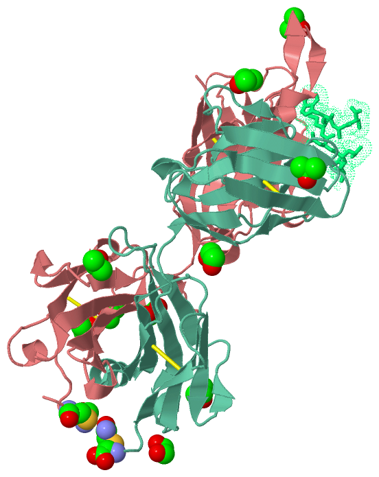
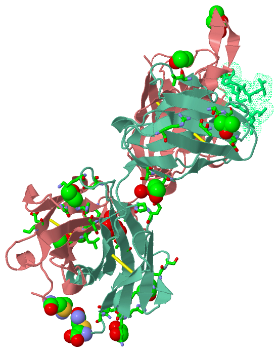
Chain H from PDB  Type:PROTEIN  Length:237
                                                                                                                                                                                                                                                                              
               SCOP domains d1tjgh1 H:1-113 Immunoglobulin heavy chain variable domain, VH                                                                      d1tjgh2 H:114-216 Immunoglobulin heavy chain gamma constant domain 1, CH1-gamma                        -- SCOP domains
               CATH domains 1tjgH01 H:1-113 Immunoglobulins                                                                                                     1tjgH02 H:114-213 Immunoglobulins                                                                   ----- CATH domains
               Pfam domains --------------------------------------------------------------------------------------------------------------------------------------------------------------------------------------------------------------------------------------------- Pfam domains
         Sec.struct. author ..eeeee...ee.....eeeeeeee.........eeeeeee......eeeeeee....eee.hhhh..eeeeee....eeeeee...hhhhheeeeeeee...eee..eeeehhhhh...ee...eeeee........eeeee..hhh.ee..eeeeeeeeeee.....eeee.hhh....eee...ee.....eeeeeeeeee.hhh.....eeeeeehhhheeeeee........ Sec.struct. author
                 SAPs(SNPs) --------------------------------------------------------------------------------------------------------------------------------------------------------------------------------------------------------------------------------------------- SAPs(SNPs)
                    PROSITE --------------------------------------------------------------------------------------------------------------------------------------------------------------------------------------------------------------------------------------------- PROSITE
                 Transcript --------------------------------------------------------------------------------------------------------------------------------------------------------------------------------------------------------------------------------------------- Transcript
                1tjg H    1 RITLKESGPPLVKPTQTLTLTCSFSGFSLSDFGVGVGWIRQPPGKALEWLAIIYSDDDKRYSPSLNTRLTITKDTSKNQVVLVMTRVSPVDTATYFCAHRRGPTTLFGVPIARGPVNAMDVWGQGITVTISSTSTKGPSVFPLAPSSKSTSGGTAALGCLVKDYFPEPVTVSWNSGALTSGVHTFPAVLQSSGLYSLSSVVTVPSSSLGTQTYICNVNHKPSNTKVDKKVEPKScDK  218
                                    10        20        30     || 38        48        58        68        78    ||| 85        95     |100E|||||||101       111       121       131       141       151       161       171       181       191       201       211    |  
                                                             35A|                                             82A||               100A|||||100J||||                                                                                                                 216-YCM
                                                              35B                                              82B|                100B|||||100K|||                                                                                                                      
                                                                                                                82C                 100C|||||100L||                                                                                                                      
                                                                                                                                     100D|||||100M|                                                                                                                      
                                                                                                                                      100E|||| 100N                                                                                                                      
                                                                                                                                       100F|||                                                                                                                           
                                                                                                                                        100G||                                                                                                                           
                                                                                                                                         100H|                                                                                                                           
                                                                                                                                          100I                                                                                                                           

Chain L from PDB  Type:PROTEIN  Length:214
                                                                                                                                                                                                                                                       
               SCOP domains d1tjgl1 L:1-107 Immunoglobulin light chain kappa variable domain, VL-kappa                                 d1tjgl2 L:108-214 Immunoglobulin light chain kappa constant domain, CL-kappa                                SCOP domains
               CATH domains 1tjgL01 L:1-107 Immunoglobulins                                                                            1tjgL02 L:108-211 Immunoglobulins                                                                       --- CATH domains
               Pfam domains ---------------------------------------------------------------------------------------------------------------------------------------------------------------------------------------------------------------------- Pfam domains
         Sec.struct. author ...eeee..eeee.....eeeeeee.......eeeeee......eeeee...ee.......eeeeee..eeeeee...hhhhh.eeeeee......ee...eeeee.......eeeee..hhhhhhhheeeeeeeeeee.....eeeeee..ee....eeeee.........eeeeeeeeeehhhhh...eeeeeee.......eeeeee.... Sec.struct. author
                 SAPs(SNPs) ---------------------------------------------------------------------------------------------------------------------------------------------------------------------------------------------------------------------- SAPs(SNPs)
                    PROSITE ---------------------------------------------------------------------------------------------------------------------------------------------------------------------------------------------------------------------- PROSITE
                 Transcript ---------------------------------------------------------------------------------------------------------------------------------------------------------------------------------------------------------------------- Transcript
                1tjg L    1 ALQLTQSPSSLSASVGDRITITCRASQGVTSALAWYRQKPGSPPQLLIYDASSLESGVPSRFSGSGSGTEFTLTISTLRPEDFATYYCQQLHFYPHTFGGGTRVDVRRTVAAPSVFIFPPSDEQLKSGTASVVCLLNNFYPREAKVQWKVDNALQSGNSQESVTEQDSKDSTYSLSSTLTLSKADYEKHKVYACEVTHQGLSSPVTKSFNRGEc  214
                                    10        20        30        40        50        60        70        80        90       100       110       120       130       140       150       160       170       180       190       200       210   |
                                                                                                                                                                                                                                               214-YCM

Chain P from PDB  Type:PROTEIN  Length:7
 aligned with Q75760_9HIV1 | Q75760 from UniProtKB/TrEMBL  Length:847

    Alignment length:7
        Q75760_9HIV1    653 ELDKWAS  659
               SCOP domains ------- SCOP domains
               CATH domains ------- CATH domains
               Pfam domains GP41-1t Pfam domains
         Sec.struct. author ....... Sec.struct. author
                 SAPs(SNPs) ------- SAPs(SNPs)
                    PROSITE ------- PROSITE
                 Transcript ------- Transcript
                1tjg P  662 ELDKWAS  668

   Legend:   → Mismatch (orange background)
  - → Gap (green background, '-', border residues have a numbering label)
    → Modified Residue (blue background, lower-case, 'x' indicates undefined single-letter code, labelled with number + name)
  x → Chemical Group (purple background, 'x', labelled with number + name, e.g. ACE or NH2)
  extra numbering lines below/above indicate numbering irregularities and modified residue names etc., number ends below/above '|'

 Classification and Annotation

(-) SCOP Domains  (4, 4)

Asymmetric/Biological Unit

(-) CATH Domains  (1, 4)

Asymmetric/Biological Unit
(-)
Class: Mainly Beta (13760)

(-) Pfam Domains  (1, 1)

Asymmetric/Biological Unit

(-) Gene Ontology  (15, 15)

Asymmetric/Biological Unit(hide GO term definitions)
Chain P   (Q75760_9HIV1 | Q75760)
molecular function
    GO:0005515    protein binding    Interacting selectively and non-covalently with any protein or protein complex (a complex of two or more proteins that may include other nonprotein molecules).
    GO:0005198    structural molecule activity    The action of a molecule that contributes to the structural integrity of a complex or its assembly within or outside a cell.
biological process
    GO:0039663    membrane fusion involved in viral entry into host cell    Merging of the virion membrane and a host membrane (host plasma membrane or host organelle membrane) that is involved in the uptake of a virus into a host cell.
    GO:0046718    viral entry into host cell    The process that occurs after viral attachment by which a virus, or viral nucleic acid, breaches the plasma membrane or cell envelope and enters the host cell. The process ends when the viral nucleic acid is released into the host cell cytoplasm.
    GO:0016032    viral process    A multi-organism process in which a virus is a participant. The other participant is the host. Includes infection of a host cell, replication of the viral genome, and assembly of progeny virus particles. In some cases the viral genetic material may integrate into the host genome and only subsequently, under particular circumstances, 'complete' its life cycle.
    GO:0019062    virion attachment to host cell    The process by which a virion protein binds to molecules on the host cellular surface or host cell surface projection.
cellular component
    GO:0044174    host cell endosome    A membrane-bounded organelle that carries materials newly ingested by endocytosis. It passes many of the materials to host cell lysosomes for degradation.
    GO:0044175    host cell endosome membrane    The lipid bilayer surrounding a host cell endosome.
    GO:0033644    host cell membrane    Double layer of lipid molecules as it encloses host cells, and, in eukaryotes, many organelles; may be a single or double lipid bilayer; also includes associated proteins. The host is defined as the larger of the organisms involved in a symbiotic interaction.
    GO:0020002    host cell plasma membrane    The plasma membrane surrounding a host cell.
    GO:0016021    integral component of membrane    The component of a membrane consisting of the gene products and protein complexes having at least some part of their peptide sequence embedded in the hydrophobic region of the membrane.
    GO:0016020    membrane    A lipid bilayer along with all the proteins and protein complexes embedded in it an attached to it.
    GO:0019031    viral envelope    The lipid bilayer of a virion that surrounds the protein capsid. May also contain glycoproteins.
    GO:0019012    virion    The complete fully infectious extracellular virus particle.
    GO:0055036    virion membrane    The lipid bilayer surrounding a virion.

 Visualization

(-) Interactive Views

Asymmetric/Biological Unit
  Complete Structure
    Jena3D(integrated viewing of ligand, site, SAP, PROSITE, SCOP information)
    WebMol | AstexViewer[tm]@PDBe
(Java Applets, require no local installation except for Java; loading may be slow)
    STRAP
(Java WebStart application, automatic local installation, requires Java; full application with system access!)
    RasMol
(require local installation)
    Molscript (VRML)
(requires installation of a VRML viewer; select preferred view via VRML and generate a mono or stereo PDF format file)
 
  Ligands, Modified Residues, Ions
    EDO  [ RasMol | Jena3D ]  +environment [ RasMol | Jena3D ]
    IPA  [ RasMol | Jena3D ]  +environment [ RasMol | Jena3D ]
    YCM  [ RasMol | Jena3D ]  +environment [ RasMol | Jena3D ]
 
  Sites
    AC1  [ RasMol ]  +environment [ RasMol ]
    AC2  [ RasMol ]  +environment [ RasMol ]
    AC3  [ RasMol ]  +environment [ RasMol ]
    AC4  [ RasMol ]  +environment [ RasMol ]
    AC5  [ RasMol ]  +environment [ RasMol ]
    AC6  [ RasMol ]  +environment [ RasMol ]
    AC7  [ RasMol ]  +environment [ RasMol ]
    AC8  [ RasMol ]  +environment [ RasMol ]
    AC9  [ RasMol ]  +environment [ RasMol ]
    BC1  [ RasMol ]  +environment [ RasMol ]
 
  Cis Peptide Bonds
    Glu H:148 - Pro H:149   [ RasMol ]  
    Phe H:146 - Pro H:147   [ RasMol ]  
    Ser L:7 - Pro L:8   [ RasMol ]  
    Tyr L:140 - Pro L:141   [ RasMol ]  
    Tyr L:94 - Pro L:95   [ RasMol ]  
 

(-) Still Images

Jmol
  protein: cartoon or spacefill or dots and stick; nucleic acid: cartoon and stick; ligands: spacefill; active site: stick
Molscript
  protein, nucleic acid: cartoon; ligands: spacefill; active site: ball and stick

 Databases and Analysis Tools

(-) Databases

Access by PDB/NDB ID
  1tjg
    Family and Domain InformationProDom | SYSTERS
    General Structural InformationGlycoscienceDB | MMDB | NDB | OCA | PDB | PDBe | PDBj | PDBsum | PDBWiki | PQS | PROTEOPEDIA
    Orientation in MembranesOPM
    Protein SurfaceSURFACE
    Secondary StructureDSSP (structure derived) | HSSP (homology derived)
    Structural GenomicsGeneCensus
    Structural NeighboursCE | VAST
    Structure ClassificationCATH | Dali | SCOP
    Validation and Original DataBMRB Data View | BMRB Restraints Grid | EDS | PROCHECK | RECOORD | WHAT_CHECK
 
Access by UniProt ID/Accession number
  Q75760_9HIV1 | Q75760
    Comparative Protein Structure ModelsModBase
    Genomic InformationEnsembl
    Protein-protein InteractionDIP
    Sequence, Family and Domain InformationInterPro | Pfam | SMART | UniProtKB/TrEMBL
 
Access by Enzyme Classificator   (EC Number)
  (no 'Enzyme Classificator' available)
    General Enzyme InformationBRENDA | EC-PDB | Enzyme | IntEnz
    PathwayKEGG | MetaCyc
 
Access by Disease Identifier   (MIM ID)
  (no 'MIM ID' available)
    Disease InformationOMIM
 
Access by GenAge ID
  (no 'GenAge ID' available)
    Age Related InformationGenAge

(-) Analysis Tools

Access by PDB/NDB ID
    Domain InformationXDom
    Interatomic Contacts of Structural UnitsCSU
    Ligand-protein ContactsLPC
    Protein CavitiescastP
    Sequence and Secondary StructurePDBCartoon
    Structure AlignmentSTRAP(Java WebStart application, automatic local installation, requires Java; full application with system access!)
    Structure and Sequence BrowserSTING
 
Access by UniProt ID/Accession number
  Q75760_9HIV1 | Q75760
    Protein Disorder PredictionDisEMBL | FoldIndex | GLOBPLOT (for more information see DisProt)

 Related Entries

(-) Entries Sharing at Least One Protein Chain (UniProt ID)

UniProtKB/Swiss-Prot
        Q75760_9HIV1 | Q757605fuu
UniProtKB/TrEMBL
        Q75760_9HIV1 | Q757602b4c 2f5b 3tyg 4jm2 5fyk

(-) Related Entries Specified in the PDB File

1tjh 2F5 FAB COMPLEXED WITH A GP41 11MER
1tji 2F5 FAB COMPLEXED WITH A GP41 17MER
2f5b 2F5 IN COMPLEX WITH ITS GP41 EPTIOPE