| molecular function |
|---|
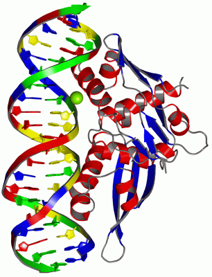
| | GO:0003677 | | DNA binding | | Any molecular function by which a gene product interacts selectively and non-covalently with DNA (deoxyribonucleic acid). |
| | GO:0003887 | | DNA-directed DNA polymerase activity | | Catalysis of the reaction: deoxynucleoside triphosphate + DNA(n) = diphosphate + DNA(n+1); the synthesis of DNA from deoxyribonucleotide triphosphates in the presence of a DNA template and a 3'hydroxyl group. |
| biological process |
|---|
| | GO:0071897 | | DNA biosynthetic process | | The cellular DNA metabolic process resulting in the formation of DNA, deoxyribonucleic acid, one of the two main types of nucleic acid, consisting of a long unbranched macromolecule formed from one or two strands of linked deoxyribonucleotides, the 3'-phosphate group of each constituent deoxyribonucleotide being joined in 3',5'-phosphodiester linkage to the 5'-hydroxyl group of the deoxyribose moiety of the next one. |
| | GO:0006260 | | DNA replication | | The cellular metabolic process in which a cell duplicates one or more molecules of DNA. DNA replication begins when specific sequences, known as origins of replication, are recognized and bound by initiation proteins, and ends when the original DNA molecule has been completely duplicated and the copies topologically separated. The unit of replication usually corresponds to the genome of the cell, an organelle, or a virus. The template for replication can either be an existing DNA molecule or RNA. |
| | GO:0006270 | | DNA replication initiation | | The process in which DNA-dependent DNA replication is started; this involves the separation of a stretch of the DNA double helix, the recruitment of DNA polymerases and the initiation of polymerase action. |
| | GO:0006276 | | plasmid maintenance | | The maintenance of the integrity of extrachromosomal plasmid DNA; includes processes that ensure plasmids are retained in the daughter cells after cell division. |
| cellular component |
|---|
| | GO:0005727 | | extrachromosomal circular DNA | | Circular DNA structures that are not part of a chromosome. |
 Description