|
|
|
|
 Description
|
|
Compounds
|
||||||||||||||||||||||||||||||||||||||||||||||||||||||||||||
Chains, Units
Summary Information (see also Sequences/Alignments below) |
Ligands, Modified Residues, Ions (0, 0)| (no "Ligand,Modified Residues,Ions" information available for 1R4G) |
Sites (0, 0)| (no "Site" information available for 1R4G) |
SS Bonds (0, 0)| (no "SS Bond" information available for 1R4G) |
Cis Peptide Bonds (0, 0)| (no "Cis Peptide Bond" information available for 1R4G) |
SAPs(SNPs)/Variants (0, 0)| (no "SAP(SNP)/Variant" information available for 1R4G) |
PROSITE Motifs (0, 0)| (no "PROSITE Motif" information available for 1R4G) |
Exons (0, 0)| (no "Exon" information available for 1R4G) |
Sequences/Alignments
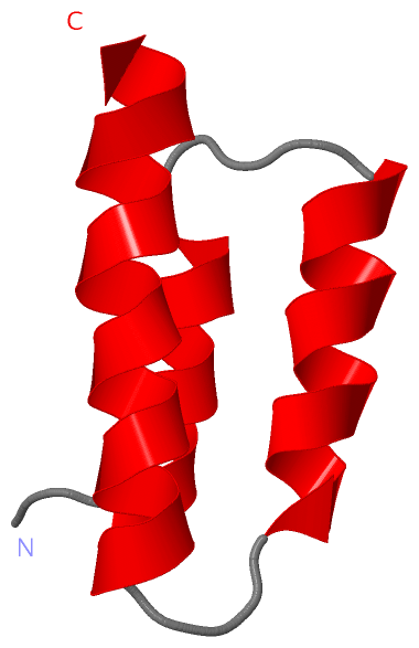
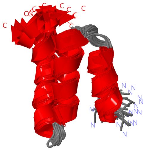
NMR StructureChain A from PDB Type:PROTEIN Length:53 aligned with PHOSP_SENDH | P04859 from UniProtKB/Swiss-Prot Length:568 Alignment length:53 525 535 545 555 565 PHOSP_SENDH 516 KPTMHSLRLVIESSPLSRAEKAAYVKSLSKCKTDQEVKAVMELVEEDIESLTN 568 SCOP domains d1r4ga_ A: RNA polymerase alpha subunit SCOP domains CATH domains ----------------------------------------------------- CATH domains Pfam domains Paramyxo_P-1r4gA01 A:516-566 -- Pfam domains SAPs(SNPs) ----------------------------------------------------- SAPs(SNPs) PROSITE ----------------------------------------------------- PROSITE Transcript ----------------------------------------------------- Transcript 1r4g A 516 KPTMHSLRLVIESSPLSRAEKAAYVKSLSKCKTDQEVKAVMELVEEDIESLTN 568 525 535 545 555 565
|
||||||||||||||||||||
SCOP Domains (1, 1)
NMR Structure
 |
CATH Domains (0, 0)| (no "CATH Domain" information available for 1R4G) |
Pfam Domains (1, 1)
NMR Structure
|
Gene Ontology (7, 7)|
NMR Structure(hide GO term definitions) Chain A (PHOSP_SENDH | P04859)
|
||||||||||||||||||||||||||||||||||||||||||||||||||||||||||||
Interactive Views
|
||||||||||||||||||||||||||||||||||||||||||||||||||||||||||||||||||||||||||||||||||||||||||||||||||||||||||||||||||||
Still Images
|
||||||||||||||||
Databases
|
||||||||||||||||||||||||||||||||||||||||||||||||||||||||||||||||||||||||||||||||||||||||||||||||||||||||||||||||||||||||||||||||||||||||||||||||||||||||||||||||
Analysis Tools
|
|||||||||||||||||||||||||||||||||||||||||||||||||||||||||||||
Entries Sharing at Least One Protein Chain (UniProt ID)
Related Entries Specified in the PDB File
|
|